- Be able to identify tissues in the nervous system (nerves, cell bodies and ganglia, and white vs. gray matter in the spinal cord, cerebellum, and cerebrum).
- Describe the organization and understand some of the basic functions of regions of the:
- spinal cord (e.g. dorsal horn, ventral horn, lateral extension of the ventral horn, and dorsal nucleus of Clarke),
- cerebellum (e.g. molecular, Purkinje, and granule cell layers and the general interactions of the cells therein)
- cerebral cortex (e.g. layers I through VI, particularly pyramidal cells of layers III and V)
- Observe ependymal cells of the choroid plexus, noting that these are the cells responsible for the production of CSF.
- Observe the 3-layered organization of the hippocampus and dentate gyrus (archicortex) as opposed to the 6-layered organization observed in other regions of the cerebral cortex (neocortex).
- Be able to identify pyramidal cells of the hippocampus and granule cells of the dentate gyrus.
Slide 065-1N spinal chord Masson cross View Virtual Slide
Slide 065-2 spinal cord lumbar H&E cross View Virtual Slide
Slide 065-1 spinal cord lumbar H&E cross View Virtual Slide
Slide 066a thoracic spinal cord thoracic spinal cord luxol blue cross View Virtual Slide
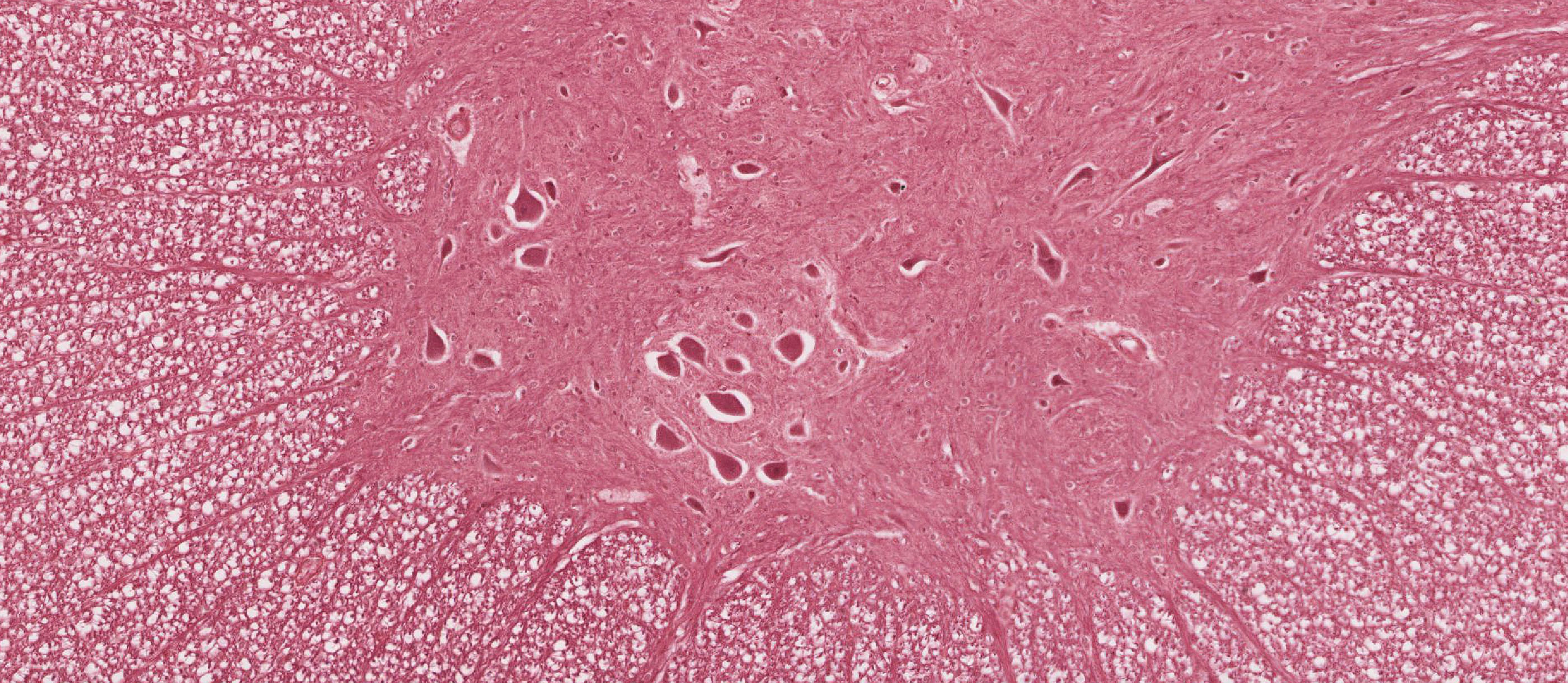
Review the organization of the spinal cord using your atlas. Examine the cross section of the lumbar spinal cord in slide 065-2. At low magnification, differentiate inner gray from outer white matter and identify dorsal and ventral horns of the gray matter. You should also identify the dorsal and ventral horns in slide 65-1N stained with Masson trichrome. In these slides, dorsal happens to be "up," but you should be able to tell dorsal and ventral horns based on morphology and the cells present rather than the orientation. The perikarya of large somatic motor neurons slide 065-2 View Image located in the ventral horn of the cord innervate the skeletal muscles of the limbs and trunk, which are embryologically derived from somites (hence, somatic muscles).
Why are perikarya of dorsal horn neurons smaller than those in the ventral horn?
Answer
Neurons in the dorsal horn are essentially interneurons that project to other regions of the CNS (e.g., motor neurons in the spinal cord or sensory input to the brain), so they have much smaller overall volume and therefore much less metabolic demand compared to motor neurons which project to target muscles that may be more than a meter away. Remember that the perikaryon is the metabolic support center for each neuron, so, therefore, motor neurons require much larger perikarya.
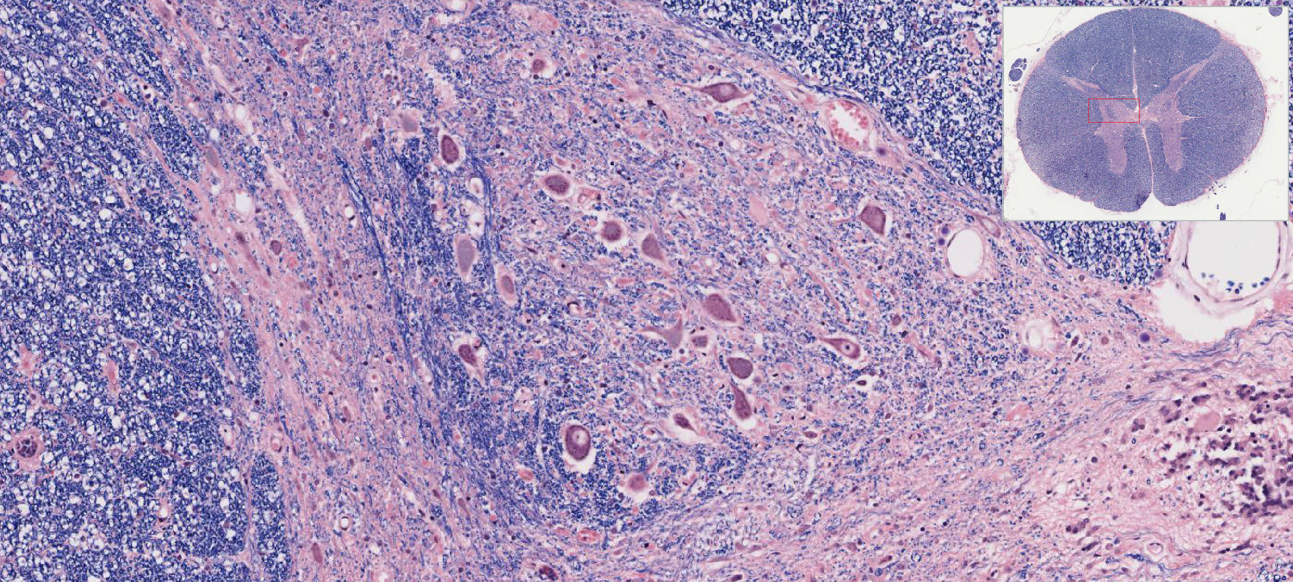
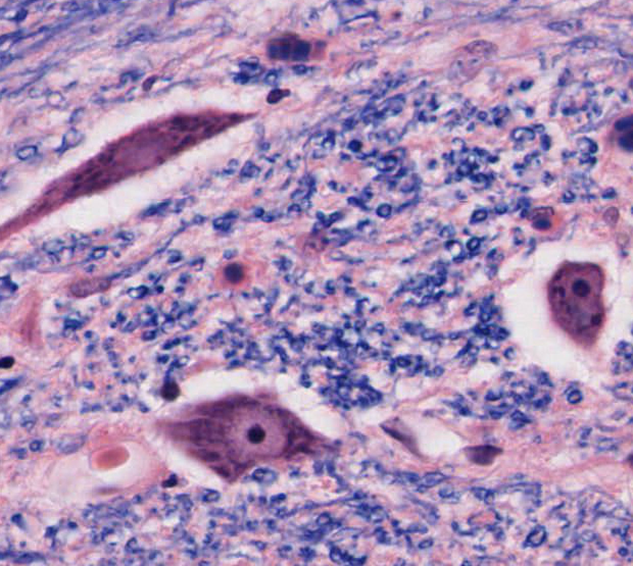
Slide 66a View Virtual Slide shows a section of thoracic spinal cord. In addition to the dorsal and ventral horns, two structures especially obvious in the thoracic cord are the dorsal nucleus of Clarke and the lateral extension of the ventral horn. The dorsal nucleus of Clarke slide 066a View Image is in the dorsal horn and contains relatively large, multipolar neurons that receive proprioceptive information from dorsal root ganglion cells that are innervated by muscle spindles in the trunk and lower limb. The cells of Clarke's nucleus then relay this information via axonal projections that extend all the way up into the cerebellum (hence the reason why the cells are so large) where it is processed to allow for coordinated movement. The lateral extension of the ventral horn slide 065-2 contains relatively large, multipolar visceral motor neurons of the intermediolateral cell column that extends from levels T1 through L2 of the spinal cord. The cells here are preganglionic sympathetic neurons whose axons terminate in either sympathetic chain ganglia or the "visceral" (or "pre-aortic") ganglia associated with the major branches of the abdominal aorta (e.g. celiac, aorticorenal, and superior/inferior mesenteric ganglia). Note that sacral levels of the cord (levels S2-4) also contain visceral motor neurons in the lateral horn, but these are parasympathetic. Many neurons in the spinal cord may appear shrunken and surrounded by an empty space due to poor fixation. Cells that are well preserved show features characteristic of most neurons: large cell body, large pale nucleus, Nissl substance, and cell processes (most of which are dendrites). The delicate meshwork of dendritic processes and nerve fibers (axons) lying between cells in the gray matter is called the neuropil. The white matter contains nerve fibers (axons) entering and exiting the gray matter, and traveling up and down the spinal cord, linking it to the brain. Nervous tissue contains two basic categories of cells: neurons and support cells (glia). Both neurons and glia have fine processes projecting from the cell body, which generally cannot be resolved in the light microscope without special staining techniques. Astrocytes in the CNS provide metabolic support for neurons and play an important role in maintaining the blood-brain barrier (see slide 13270 astrocytes View Virtual Slide). Oligodendrocytes (another type of glial cell) are responsible for the myelination of CNS axons. Recall thatSchwann cells are the glial cells responsible for myelination in the peripheral nervous system. Myelin is lipid-rich, and on gross inspection appears white. Thus, in the 'white matter' of the brain and spinal cord, myelinated axons are the predominant neuronal cell component and most of the the nuclei that you see in white matter are primarily of glial cells. The ‘gray matter’ contains relatively more neuronal and glial perikarya, as well as non-myelinated (e.g. dendritic) processes. The other major glial cell type you should know about are microglia which are small cells derived from blood monocytes. They are considered part of the mononuclear phagocytic system and will proliferate and become actively phagocytic in regions of injury and/or inflammation. Because of the difficulty of discerning each glial cell type by routine light microscopy, you will not be required to identify glial cells in HE-stained sections by light microscopy, but you should be aware of their functions.
Neurons are characterized by a large cell body or perikaryon containing a large, pale (active, euchromatic) nucleus with a prominentnucleolus. Scattered in the cytoplasm are the characteristic clusters of ribosomes and rough ER termed Nissl bodies or Nissl substance slide 066a View Image. One or more cell processes may also be seen emerging from the neuronal perikaryon. Review diagrams illustrating the morphology of neurons in your textbooks. The dendrites receive neural input from other neurons viasynapses (or they are specialized to receive sensory stimuli), and they transmit neural information toward the perikaryon (Law of Dynamic Polarization). A single axon (often called a nerve fiber) leaves the perikaryon and transmits neural signals to other neurons or to the effectororgan (e.g., skeletal muscles) via synapses.
Glial Cells
Slide NP004N hippocampal region, coronal section, luxol blue View Virtual Slide
Slide 13270 astrocytes, Gold-staining View Virtual Slide
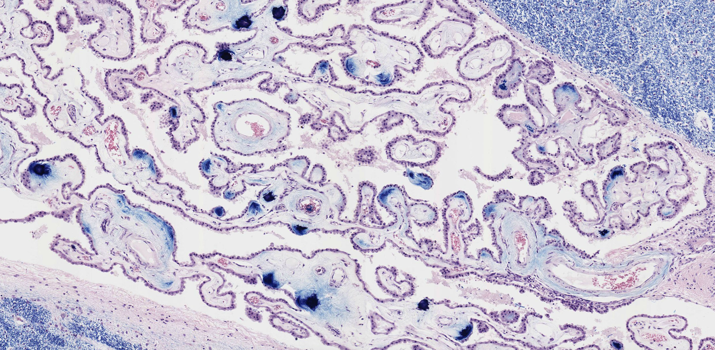
Many types of glial cells require special histological stains and can’t be unambiguously identified in regular H&E-stained histological slides. Ependymal cells, which are uniquely located lining the ventricles of the brain the central canal of the spinal cord, are one notable exception. Use the virtual slide of the hippocampal region to study the ependymal cell lining of the choroid plexus. Also note these columnar cells lining the ventricles of the brain. Slide 13270 astrocytes, Gold-staining View Virtual Slide Go to a lighter stained area of the slide, which is in focus, and look for typical star-shaped cells, which represent astrocytes. Many of these astrocytes send out processes that contact and wrap around nearby capillaries, which are also clearly recognizable as tube-shaped segments.
Slide 077 20X Cerebellum white and grey matter H&E View Virtual Slide
Slide 077 40X Cerebellum molecular layer, Purkinje cell bodies H&E View Virtual Slide
Slide 077a Cerebellum luxol blue cross View Virtual Slide
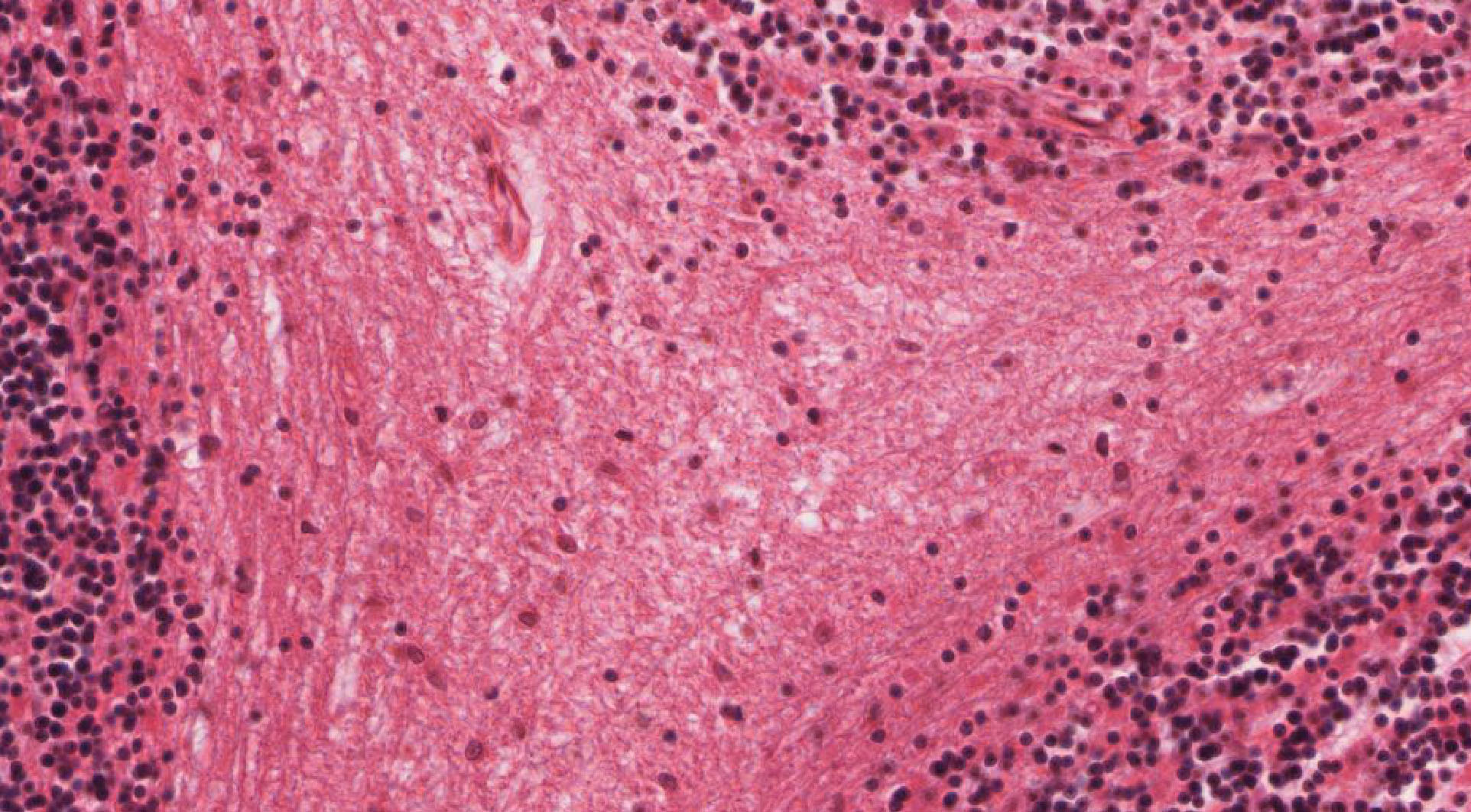
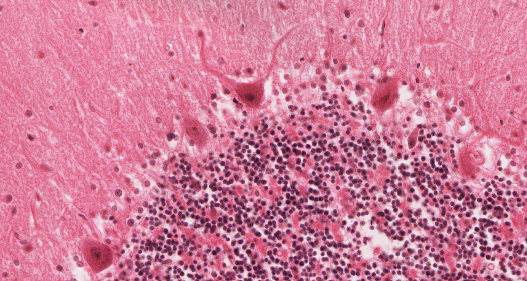
Using slide 77, determine that the cerebellar cortex is organized into an outer molecular layer slide 077 View Image containing basket and stellate cells (not distinguishable by routine light microscopy) as well as axons of granule cells found in the deeper, highly cellular granule layer slide 077 View Image. Still deeper is the white matter slide 077 View Image of the cerebellum, which contains nerve fibers, neuroglial cells, small blood vessels, but no neuronal cell bodies. Examine the boundary between molecular and granule cell layers. Here you will see the Purkinje cell bodies slide 077 View Image. In these slides you will not be able to discern the amazing dendritic tree that extends from the Purkinje cell bodies into the molecular layer, nor will you be able to see their axons, which extend down through the granular layer into deeper parts of the cerebellum. The dendritic tree and axon of each Purkinje cell can only be seen in thicker sections stained with special silver stains. Most of the nuclei visible in the granular layer belong to very small neurons, granule cells, which participate in the extensive intercommunication involved in the cerebellum’s role in balance and coordination.
Slide 076 cerebrum cerebrum luxol blue cross View Virtual Slide
Slide 076b cerebrum TB&E View Virtual Slide
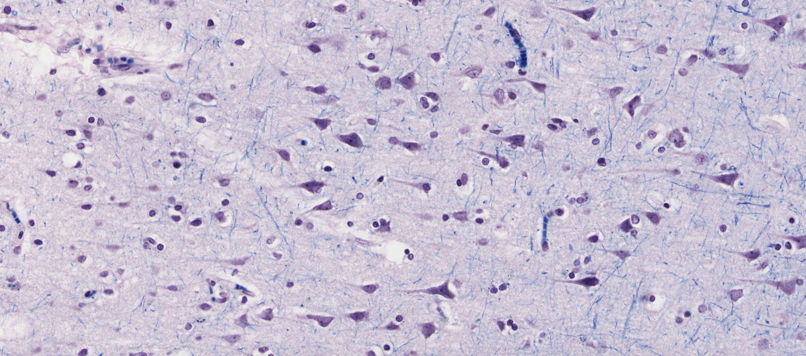
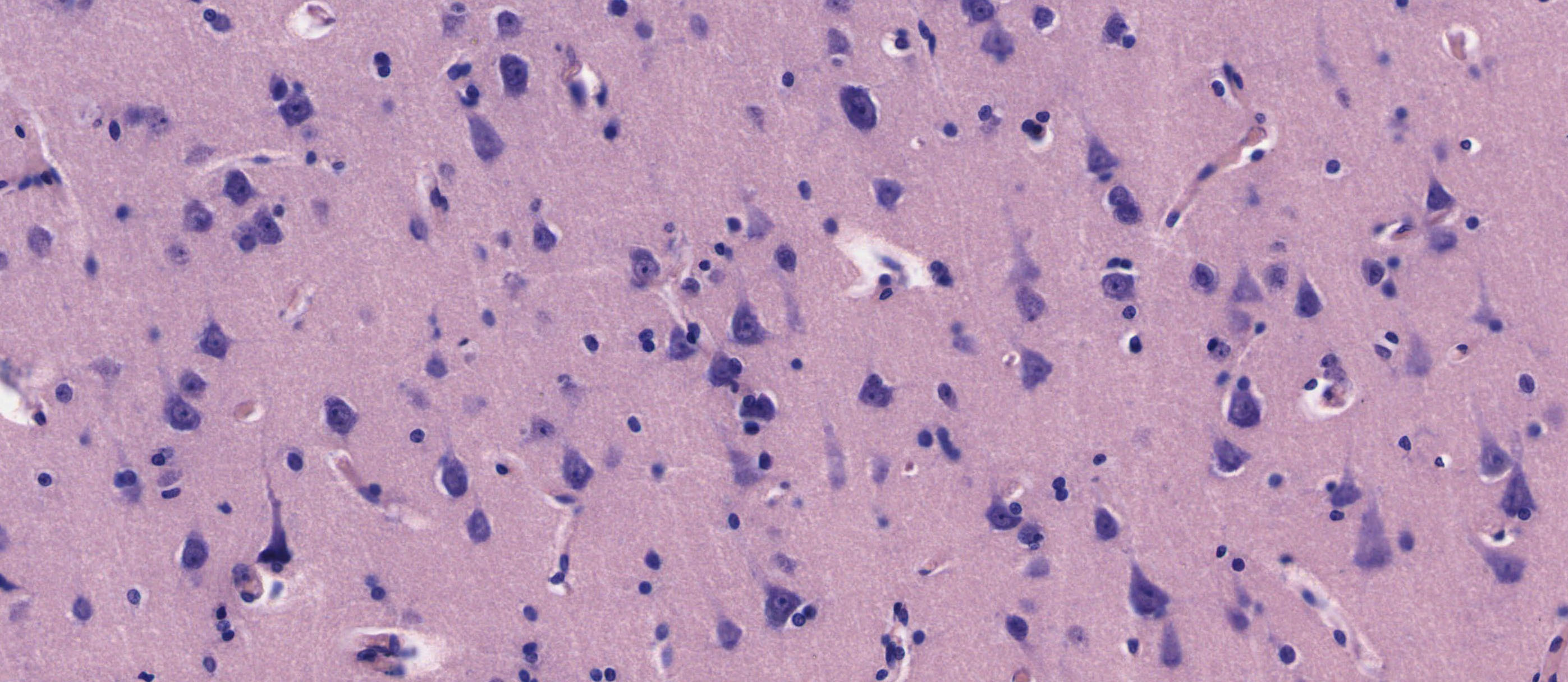
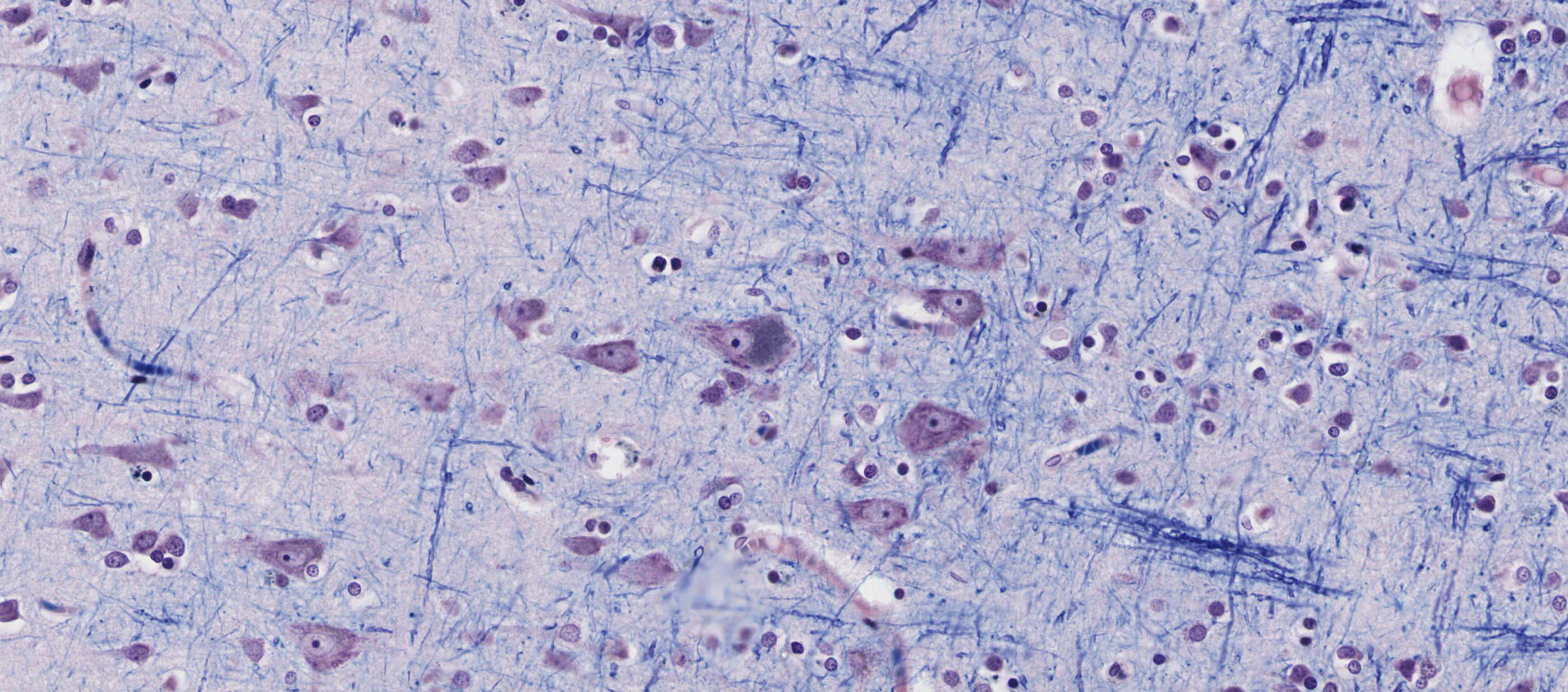
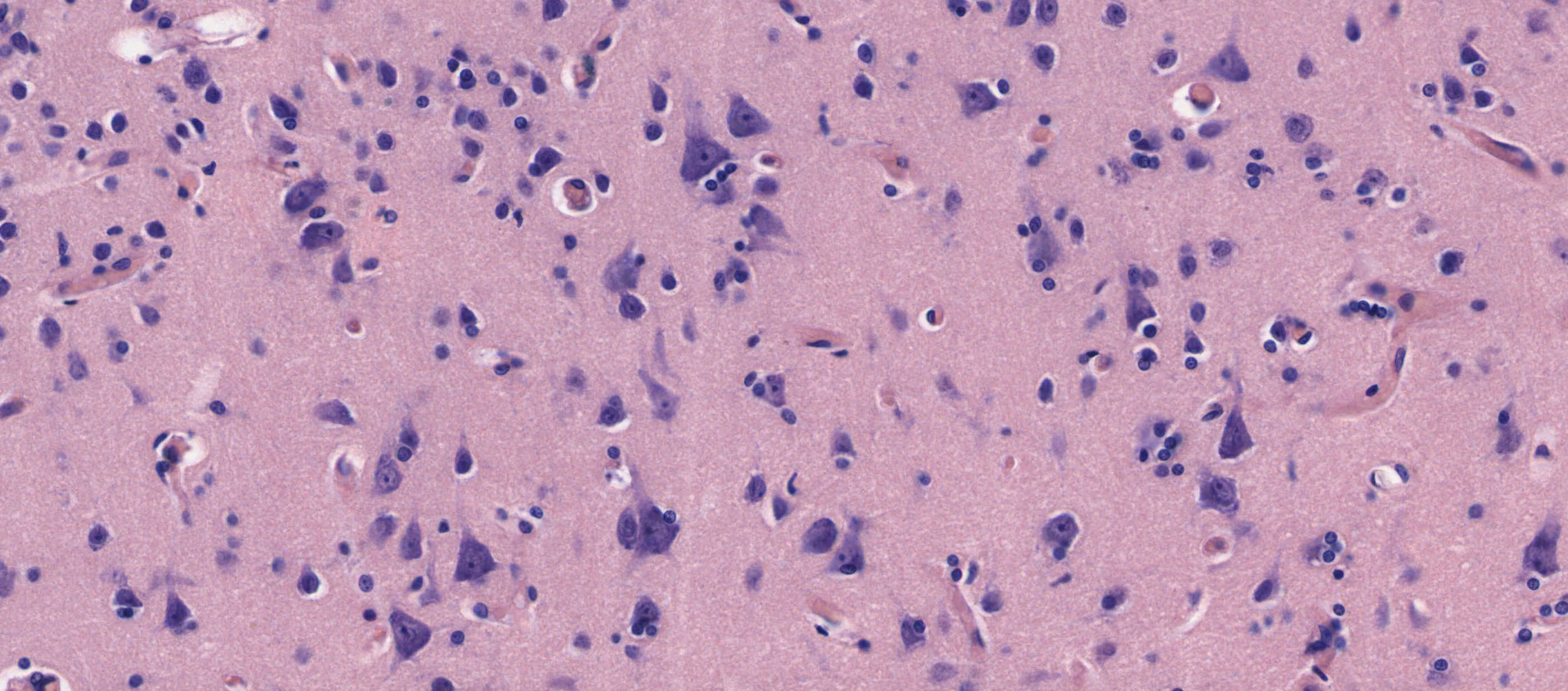
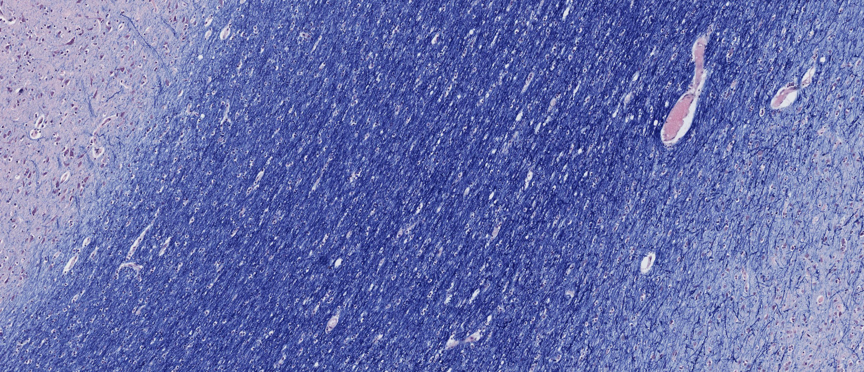
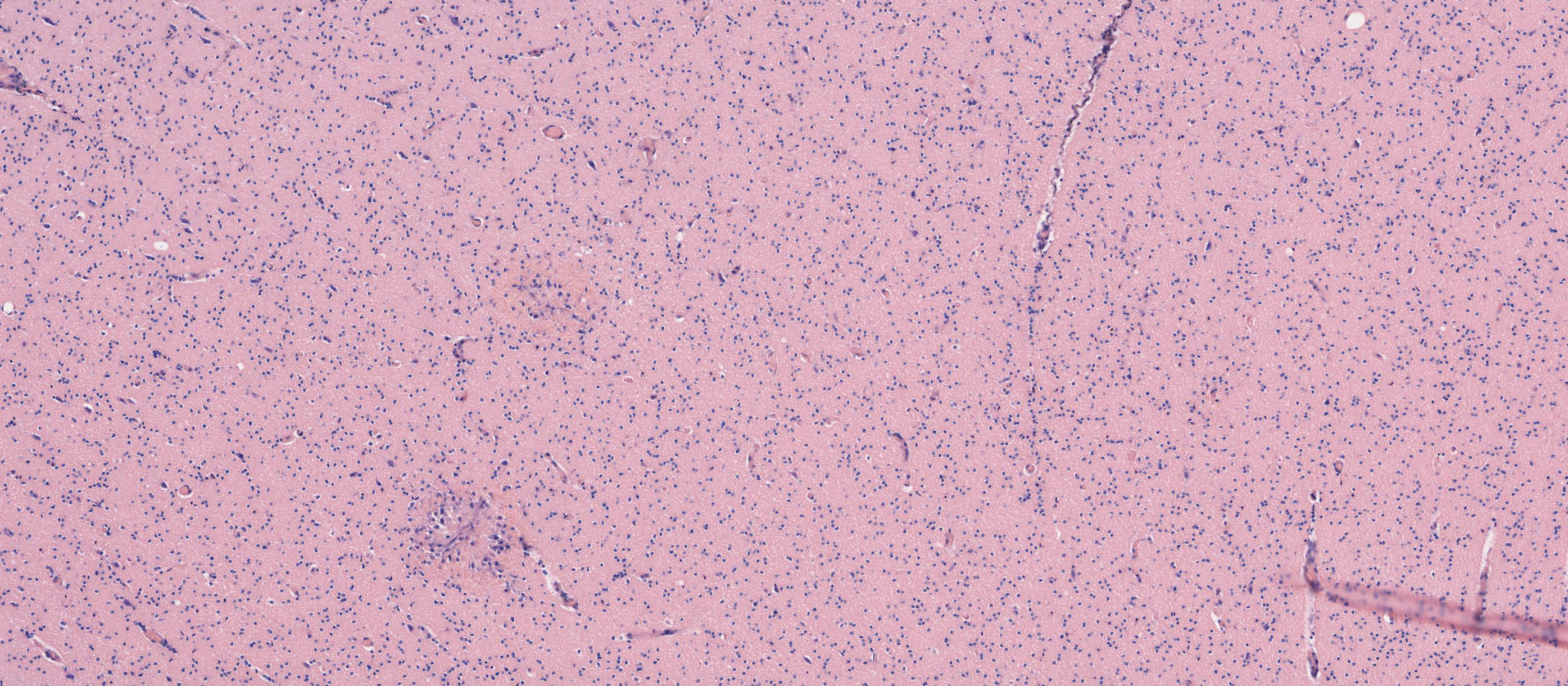
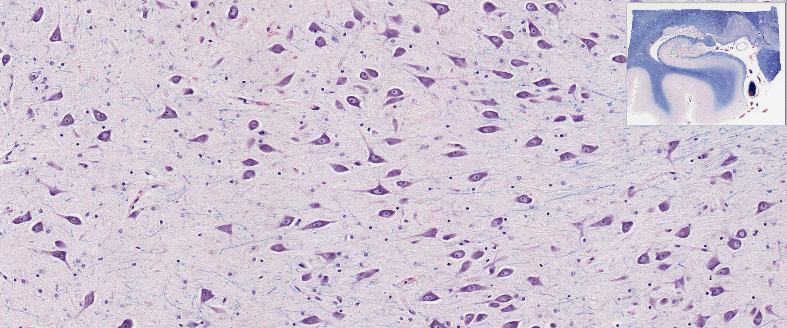
Unlike the highly organized cerebellar cortex, the cerebral cortex appears to be less well-organized when viewed with the light microscope. Nonetheless, it is loosely stratified into layers containing scattered nuclei of both neurons and glial cells. Examine the layered organization of the cerebral cortex using slide 76 stained with luxol blue/cresyl violet [orientation] (which stains white matter tracts and cell bodies) or toluidine blue and eosin [orientation] (TB&E, toluidine blue stains the nuclei and RER of cells whereas eosin stains membranes and axon tracts). Typically one or more sulci (infoldings) will extend inward from one edge of the section. Examine the gray matter on each side of the sulcus using first low and then high power. Neurons of the cerebral cortex are of varying shapes and sizes, but the most obvious arepyramidal cells. As the name implies, the cell body is shaped somewhat like a pyramid, with a large, branching dendrite extending from the apex of the pyramid toward the cortical surface, and with an axon extending downward from the base of the pyramid. In addition to pyramidal cells, other nuclei seen in these sections may belong to other neurons or to glial cells also present in the cortex. You may be able to see subtle differences in the distribution of cell types in rather loosely demarcated layers. There are 6 classically recognized layers of the cortex:
![[orientation]](/sites/default/files/images/slides/8CNS.jpg)
![[orientation]](/sites/default/files/images/slides/9CNS.jpg)
- Outer plexiform (molecular) layer: sparse neurons and glia
- Outer granular layer: small pyramidal and stellate neurons
- Outer pyramidal layer: moderate sized pyramidal neurons (should be able to see these in either luxol blue-stained slide 076 View Image or TB&E-stained slide 076b View Image sections)
- Inner granular layer: densely packed stellate neurons (usually the numerous processes aren’t visible, but there are lots of nuclei reflecting the cell density)
- Ganglionic or inner pyramidal layer: large pyramidal neurons (should be able to see these in either luxol blue-stained slide 076 View Image or TB&E-stained slide 076b View Image sections)
- Multiform cell layer: mixture of small pyramidal and stellate neurons
Pyramidal cells in layers III and V tend to be larger because their axons contribute to efferent projections that extend to other regions of the CNS –pyramidal neurons in layer V of motor cortices send projections all the way down to motor neurons in the spinal cord! Deep to the gray matter of the cerebral cortex is the white matter that conveys myelinated fibers between different parts of the cortex and other regions of the CNS. Be sure you identify the white matter in both luxol blue-stained slide 076 View Image and TB&E-stained #076b View Image sections, as it will appear differently in these two stains. Review the organization of gray and white matter in cerebral cortex vs. spinal cord.
Slide NP004N hippocampal region coronal section luxol blue View Virtual Slide [orientation]
![[orientation]](/sites/default/files/images/slides/16CNS.jpg)
This coronal section includes the hippocampus (hippocampus = sea horse), dentate gyrus, and adjacent temporal lobe gyrus (entorhinal cortex). Above the temporal (ventral or inferior) horn of the lateral ventricle the lateral geniculate nucleus is present. Lateral to this structure is the tail of the caudate. The medial surface of the section is the posterior portion of the thalamus and a small portion of the cerebral peduncle. Look at the margins of the ventricle at higher magnification and note that it is entirely lined by ependymal cells. Just medial (to the right) of the tail of the caudate, note the choroid plexus slide NP004N View Image, which consists of highly convoluted and vascularized villi covered by ependymal cells which are specialized for the production of cerebrospinal fluid, or CSF.
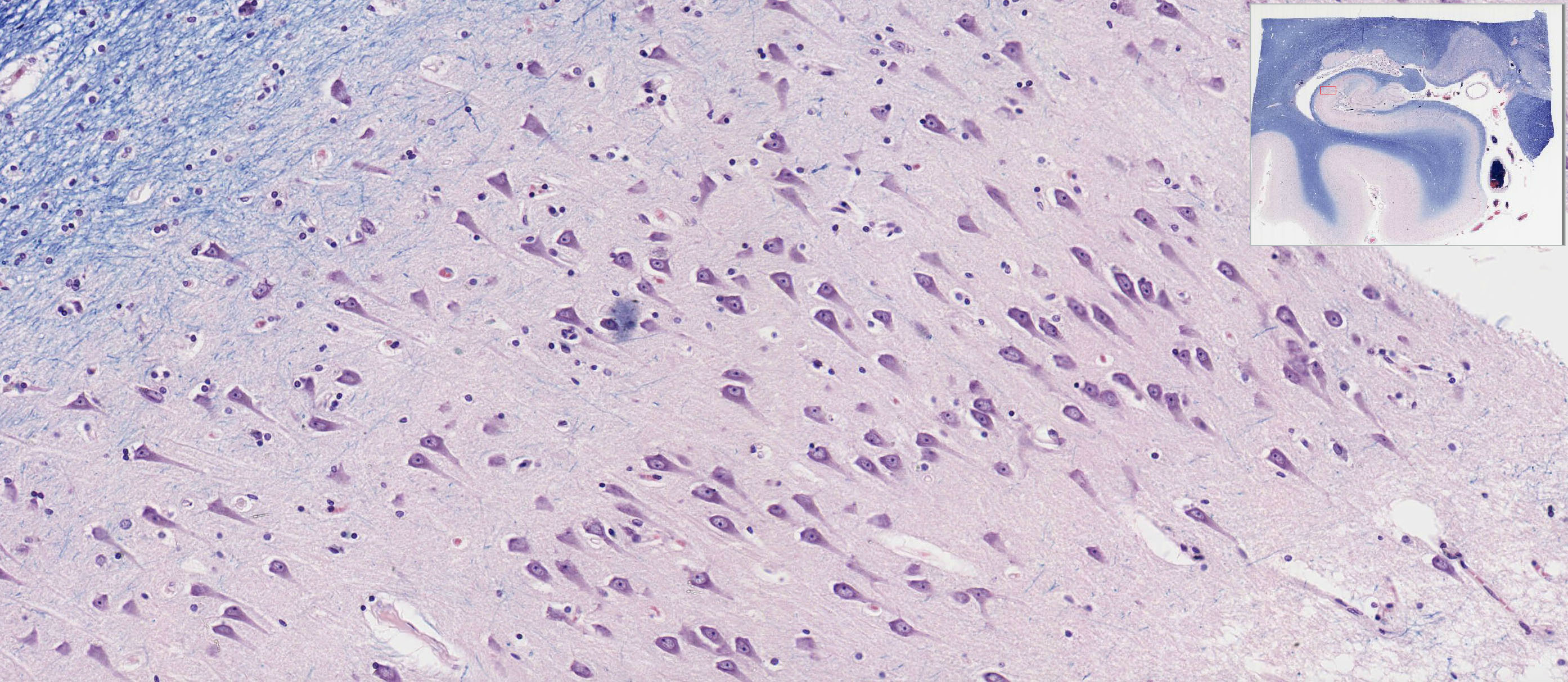
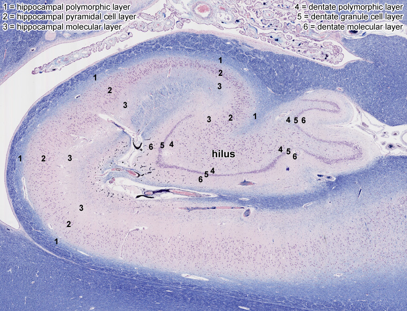
Later in this sequence, you will learn how the hippocampus and dentate gyrus function in what is known as the "limbic system" to integrate inputs from many parts of the nervous system into complicated behaviors such as learning, memory, and social interaction. For now, focus just on the morphology of these regions and observe the presence of three distinct layers rather than the six layers found in the cerebral cortex (evolutionarily speaking, the three-layered organization is considered to be "older," so this type of cortex is also known as "archicortex" whereas the "newer" six-layered cerebral cortex is "neocortex"). In the hippocampus orientation Image, observe:
- ("1" in the orientation figure) a polymorphic layer containing many nerve fibers and small cell bodies of interneurons,
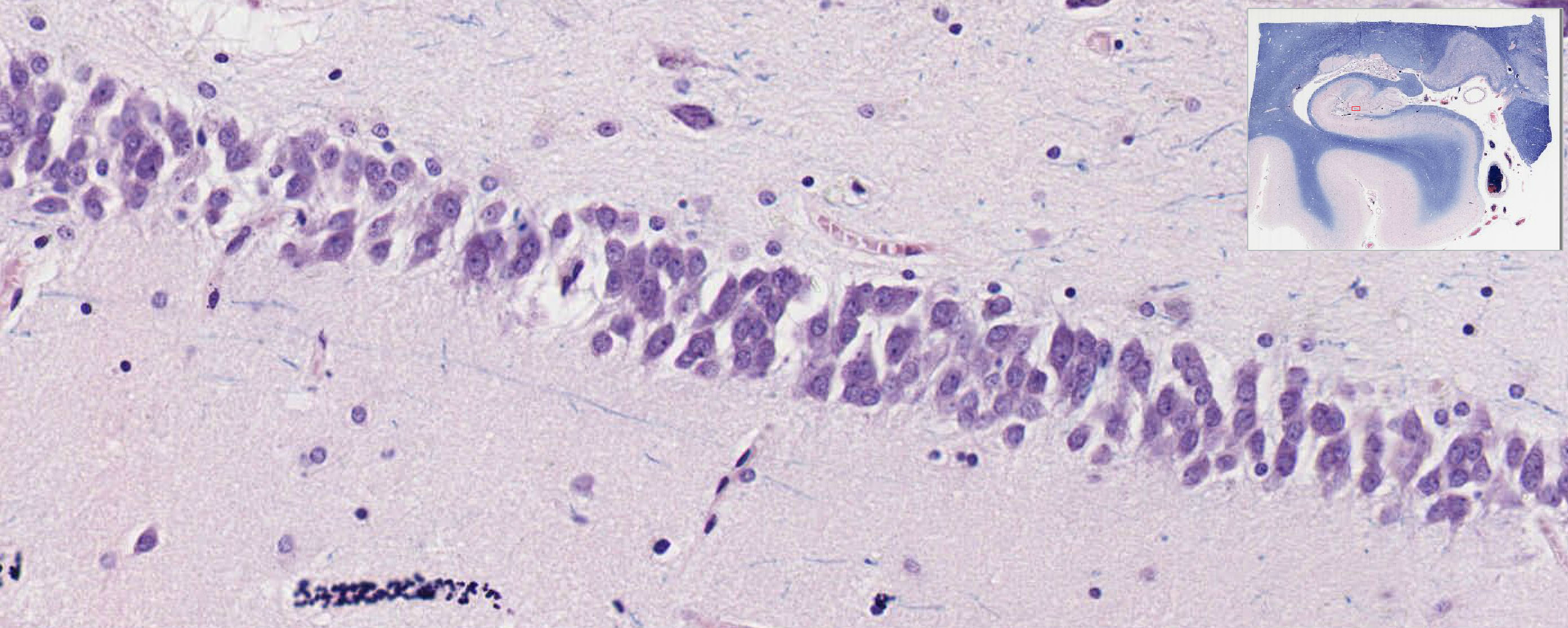
- ("2" in the orientation figure) a middle pyramidal cell layer containing hippocampal pyramidal cells slide NP004N View Image, and
- ("3" in the orientation figure) a molecular layer containing dendrites of the pyramidal cells.
In the dentate gyrus orientation Image, observe:
- ("4" in the orientation figure) a polymorphic layer containing nerve fibers (known as "mossy fibers") and cell bodies of interneurons,
- ("5" in the orientation figure) a middle granule cell layer containing the round, neuronal cell bodies of dentate granule cells slide NP004N View Image, and
- ("6" in the orientation figure) a molecular layer containing dendrites of the granule cells.
The "hilus" is the region where the head of hippocampus abuts the dentate gyrus. The multipolar neurons in this area are known as "mossy cells" slide NP004N View Image and they primarily receive input from mossy fibers of the granule cells of the dentate gyrus and then relay those signals back to other cells in the dentate. In terms of clinical significance, the pyramidal cells of the hippocampus are particularly vulnerable to damage in severe circulatory failure and by anoxia of persistent severe seizures. You may see small calcific bodies in part of the hippocampus, which occur as a normal part of the aging process. Calcific bodies are present in the choroid plexus, another common site of accumulation as the years pass.
48 Spinal Cord White Matter (Spinal Cord) View Virtual EM Slide In this field you see several oligodendrocytes, the cells that make myelin in the CNS, surrounded by numerous myelinated axons of various size, cut in cross section.
49 Motor nerve cell - Ventral Horn of Rabbit Spinal Cord, Multipolar Motor Neuron Cell Body View Virtual EM Slide Motor Neuron Cell Body. In this electron micrograph, note some of the features you saw in ventral horn motor neurons with the light microscope, such as the large, pale nucleus, prominent nucleolus, Nissl bodies, dendrites and axon. Adjacent to the neuron, note myelinated axons of various sizes and also that there are no spaces between cell processes. All spaces are occupied either by the processes of neurons or glia or by capillaries (these capillaries are somewhat swollen here because the tissue was fixed by perfusion).
Click on a question to reveal the answer.
Why are perikarya of dorsal horn neurons smaller than those in the ventral horn?
Neurons in the dorsal horn are essentially interneurons that project to other regions of the CNS (e.g. motor neurons in the spinal cord or sensory input to the brain), so they have much smaller overall volume and therefore much less metabolic demand compared to motor neurons which project to target muscles that may be more than a meter away. Remember that the perikaryon is the metabolic support center for each neuron, so, therefore, motor neurons require much larger perikarya.
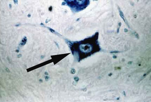
1. The arrow indicates a morphological type of neuron. With one exception, neurons such as this (though not necessarily this large) are found in ALL areas of the nervous system. Name this exception.
View Image
- Cerebellar cortex
- Cerebral cortex
- Gray matter of the spinal cord
- Dorsal root ganglia
- Peripheral autonomic ganglia
Answer
Correct answer 4. The neuron shown is a multipolar neurons. The neuronal type is found throughout the entire human nervous system except in dorsal root/sensory ganglia. These exclusively contain neurons with a pseudounipolar neuronal arrangement.
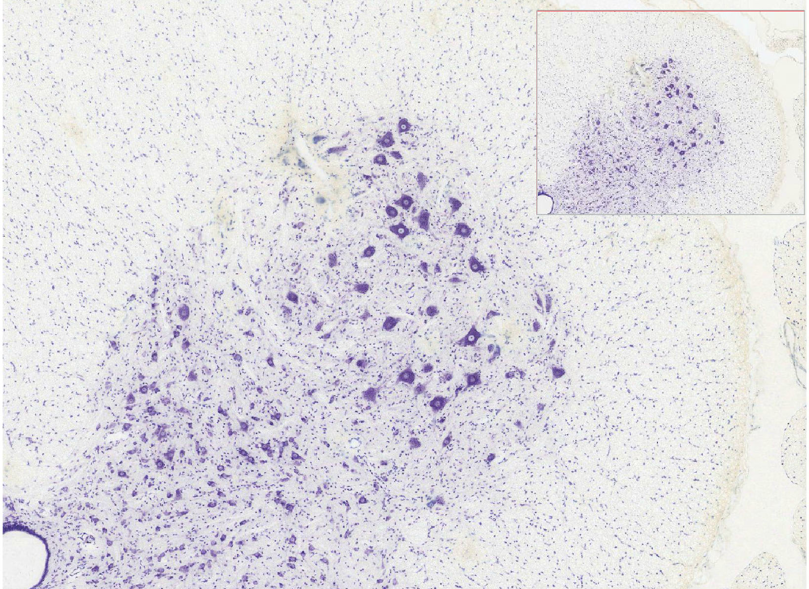
2. Name the part of the central nervous system that is displayed in this tissue section.
View Image
- Dorsal spinal cord
- Ventral spinal cord
- Cerebellum
- Hippocampus
- Dentate gyrus
Answer
Correct answer 2. The ventral spinal cord. The organization and morphology of the cells shown is found ONLY in the ventral spinal cord.
3. Which of the following cell types contributes to maintenance of the blood-brain barrier?
- Astrocytes
- Oligodendrocytes
- Microglia
- Ependymal cells of the choroid plexus
- Neurons
- NONE of the above
Answer
Correct answer 1. Astrocytes - Technically, the junctions between endothelial cells constitute the actual "barrier." However, the endothelial cells maintain these junctions in response to signals (via foot processes) from ASTROCYTES.
4. Name the function of the cell marked by the black arrow.
View Low Mag Image
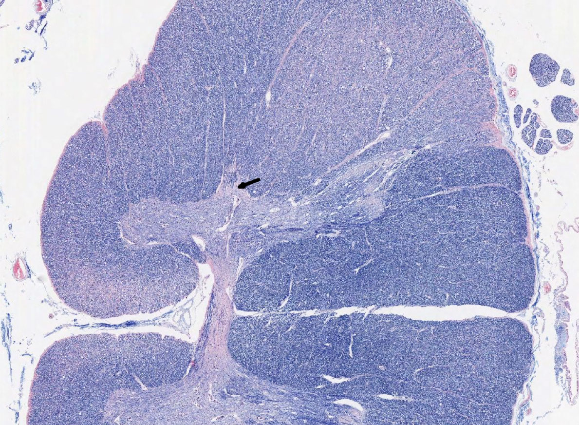
View High Mag Image
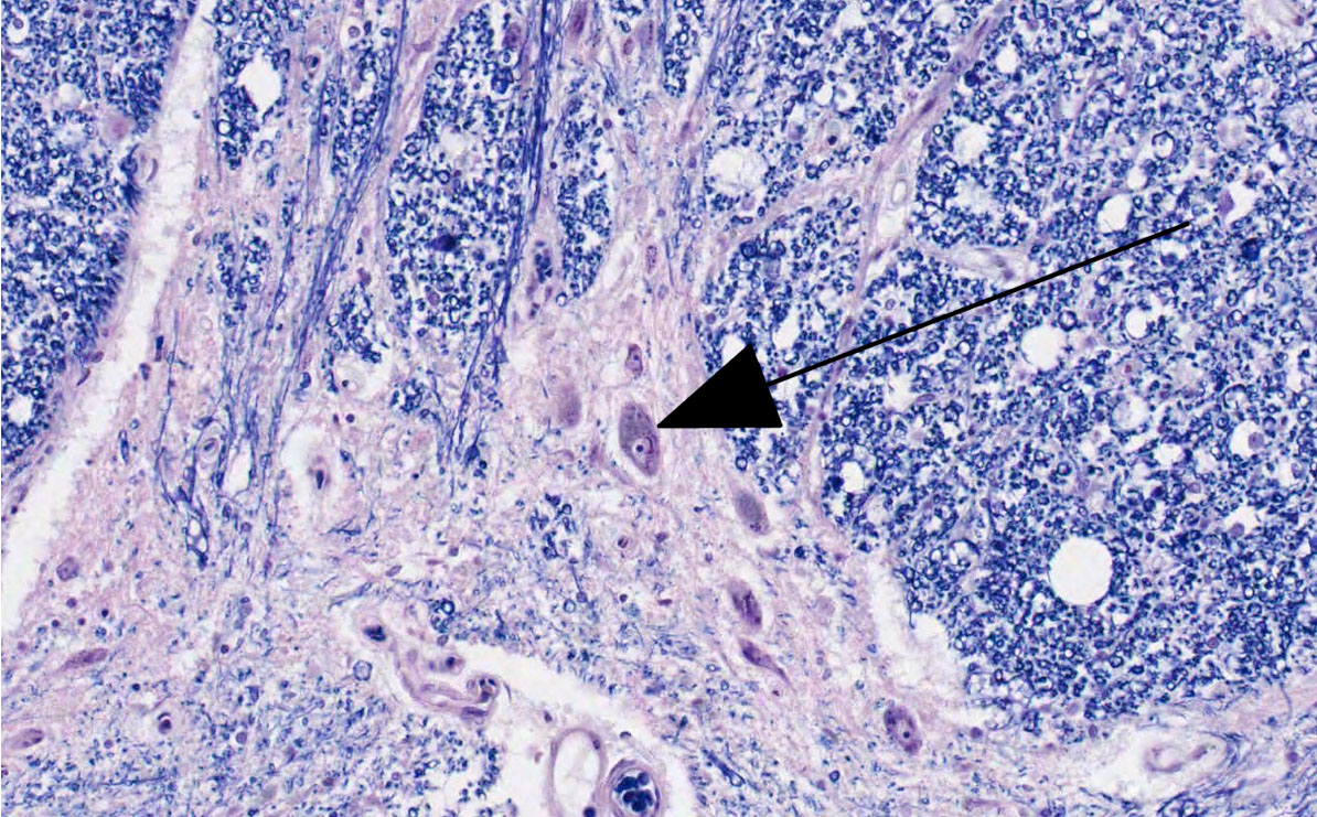
- It provides preganglionic visceral motor output to sympathetic ganglia.
- It provides somatic motor output to skeletal muscles.
- It relays sensory input to other neurons in the spinal cord.
- It relays sensory input to cerebral cortex
- It relays sensory input to cerebellar cortex.
Answer
Correct answer 1. This neuron provides preganglionic visceral motor output to sympathetic ganglia - Even though the cord is oriented "sideways," you should still be able to identify this cell as being in the intermediolateral cell column in the lateral extension of the ventral horn where pregagnglionic sympathetic visceral motor neurons are found.