Liver and Gallbladder
- Discuss the blood supply of the liver
- Define the concept of the classic liver lobule and recognize it in histological tissue sections
- Learn about the structure of portal triads and identify its components
- Understand the structure of hepatic cords and liver sinusoids
- Learn about and identify the cells of the liver tissue: hepatocytes, Kupffer cells, endothelial cells and Ito cells
- Discuss the functions and ultrastructural features of hepatocytes
- Understand the concept of the Acinus of Rappaport
- Discuss the production of bile and the cellular structures involved
- Study the histological features of the gallbladder
Slide 001 Liver (monkey) H&E View Virtual Slide
Task:
- Overview of liver organization
Using the low power objective observe numerous small, pale spots in the parenchyma, most of which are either central veins or small branches of portal veins (in portal canals). There may be a few larger channels, which are larger veins either entering or leaving this region of the liver. Try to identify classic liver lobules vs. portal lobules vs. acini of Rappaport.
Slide 194 Liver, gall bladder H&E View Virtual Slide
Slide 198 Liver, silver stain showing the collagen III reticular fibers in the space of Disse View Virtual Slide
Tasks:
- Identify central veins and portal triads; identify components of portal triads (the links below show these in slide 001, but you should try to find them on your own in slide 194)
- Follow the flow of blood between hepatic cords
- Identify Kupffer cells
- Look for the endothelial lining of the liver sinusoids
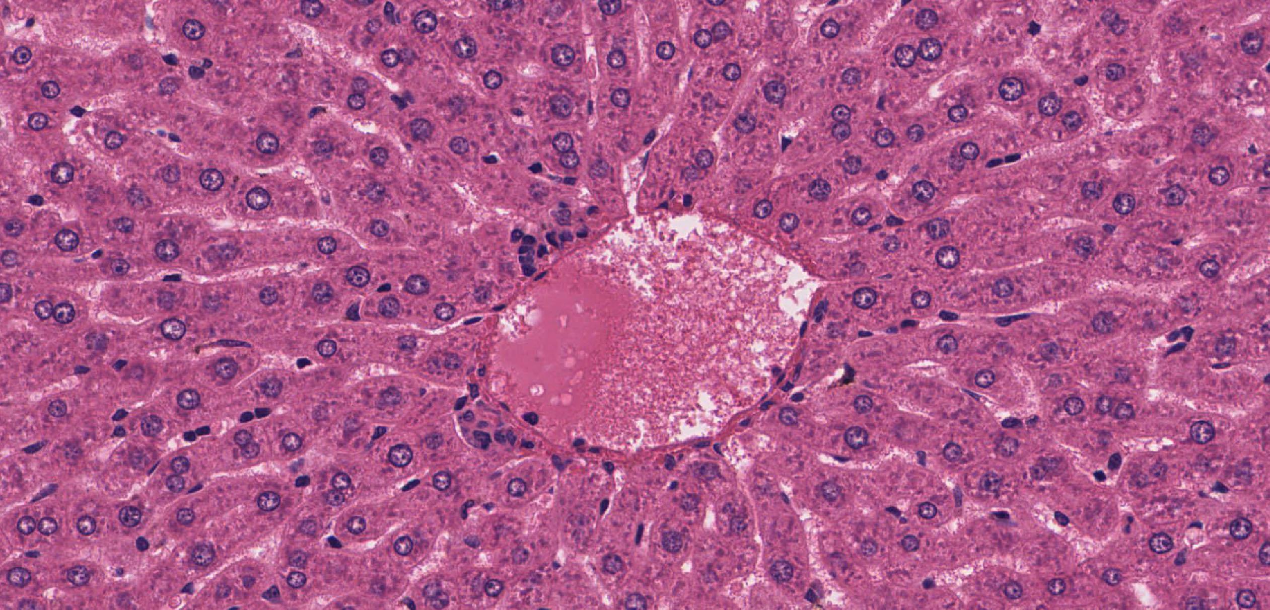
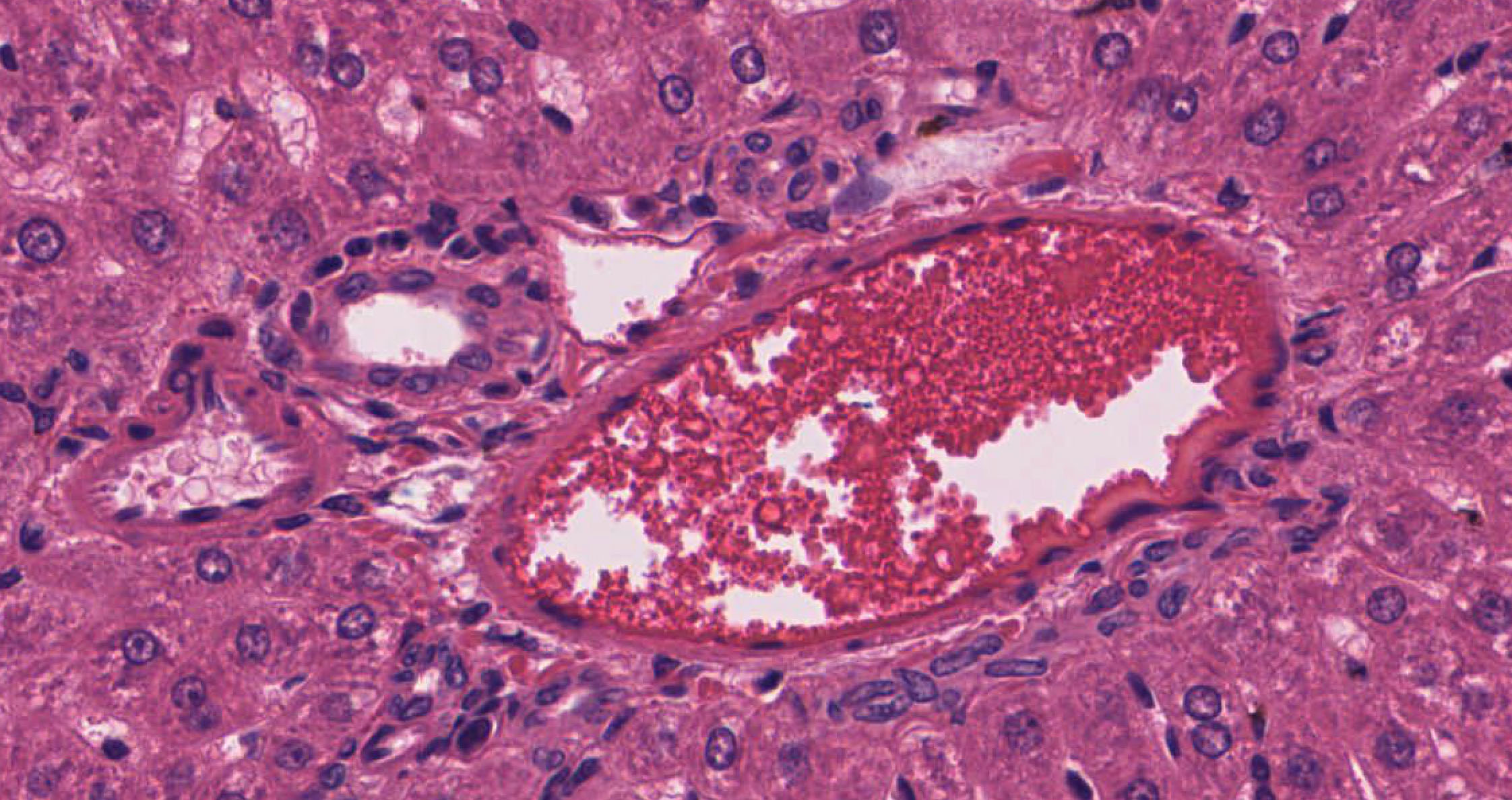
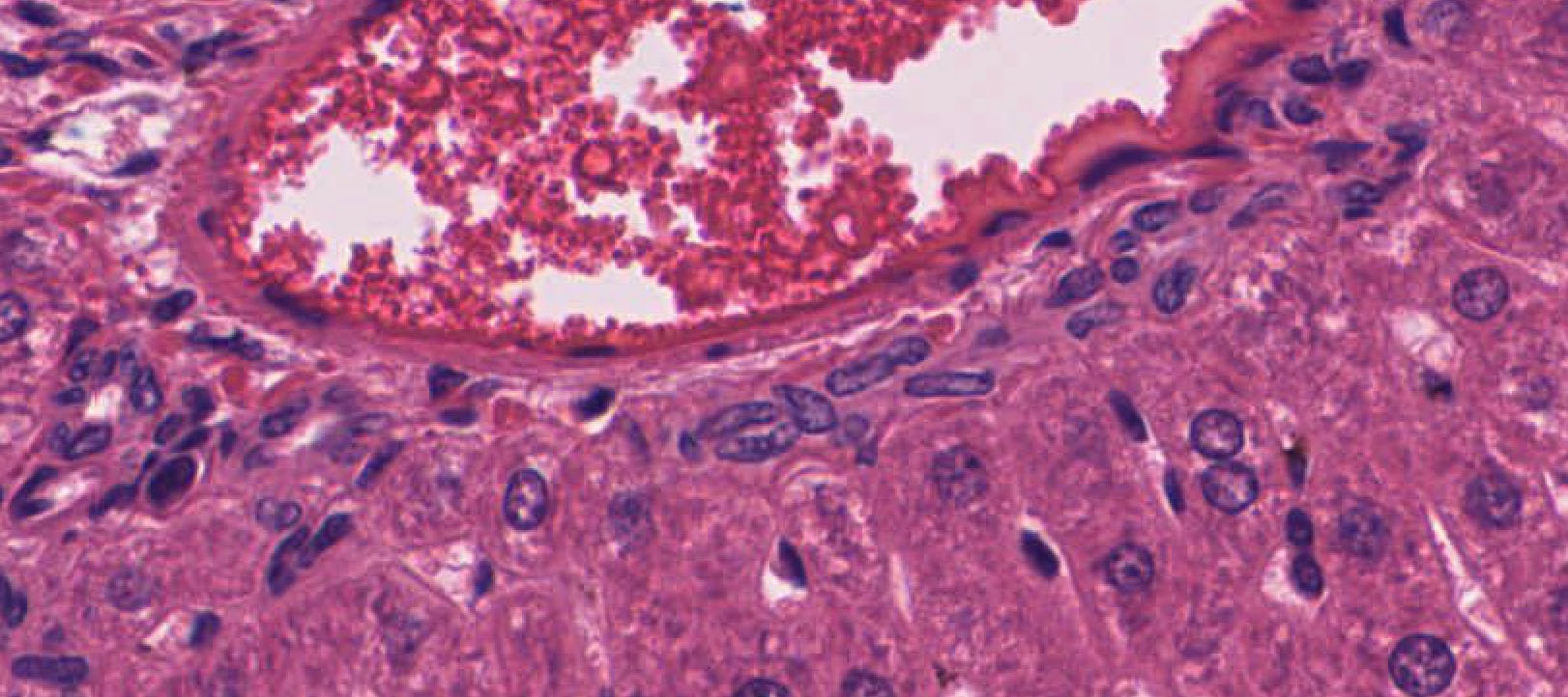
The central veins slide 001 central veins, terminal hepatic venules View Image (also referred to as terminal hepatic venules) are surrounded intimately by hepatocytes similar to those that make up the bulk of the liver tissue. Portal veins slide 001 portal vein View Image at medium power appear in section as a circle of rather prominent nuclei. In small branches of the hepatic artery slide 001 hepatic artery View Image you will see primarily the ring of smooth muscle that makes up their wall. The three components together (portal vein, hepatic artery, bile duct) constitute a portal triad. Look for good examples of portal canals where all three components are seen well. Keep in mind that these structures twist and turn so there may be more than one cross section of a bile duct, artery, or vein, so it's not always a "triad" of structures that you'll see in the portal canal. Now, see if you can define a classic liver lobule at low power.
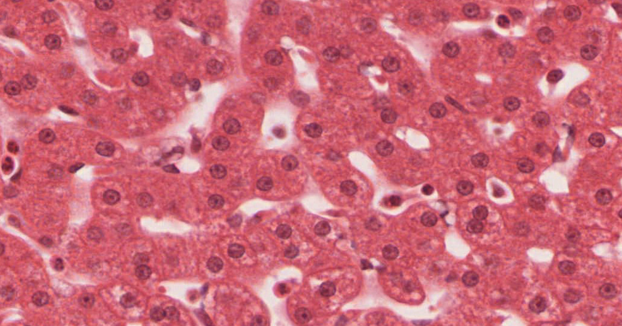
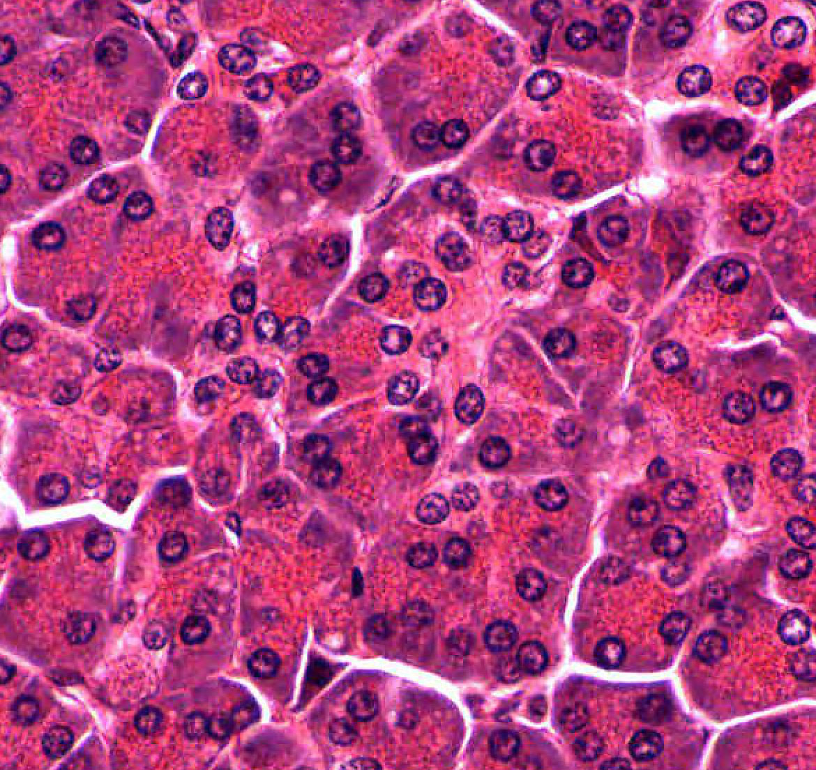
In the hepatic parenchymal tissue, note the plates of hepatocytes (the arrangement of these cells in plates is not always clear, due to plane of section and the frequent interconnections of plates). Occasional hepatocytes are binucleate. Between the plates of hepatocytes are intervening sinusoids lined by a thin endothelium. Larger eosinophilic cells lining the sinusoids are mostly Kupffer cells slide 194 Kupffer cells View Image (a type of macrophage, part of the mononuclear phagocyte system). Look for Kupffer cells using slide 194 as these cells are not readily recognized in slide 001. You should be able to distinguish Kupffer cells from endothelial lining cells.
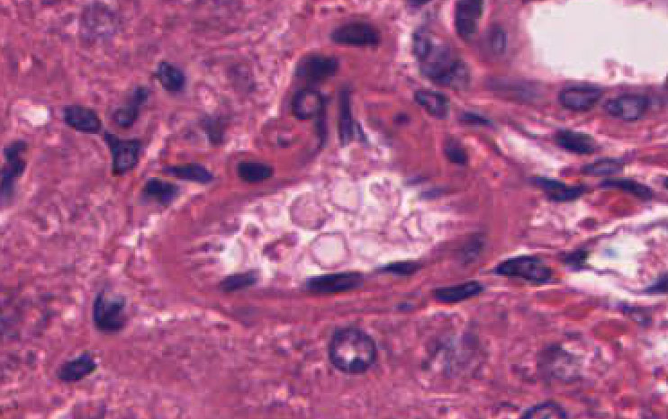
The space between the endothelial cells and hepatocytes is called the “space of Disse” and is somewhat artificially enlarged in conventional sections. The reticular fibers in this space of Disse are visualized in silver-stained slide 198. Remember that blood flows from the portal veins and hepatic arteries (of the portal canals) through the sinusoids to the central veins. A classical liver lobule has a central vein in its center and has several portal triads at its periphery. Bile flows through the bile canaliculi (too small to see) to the canals of Hering to bile ducts in portal canals, to hepatic ducts of increasing sizes and to the common hepatic duct, eventually to be emptied into the duodenum via the common bile duct. If you really want to find a canal of Hering, look for a line of low cuboidal cells slide 001 canal of Hering View Image immediately adjacent to a portal canal --the canal of Hering connects canaliculi to the bile duct.
This portal inflow system can be distinguished from the portal outflow system which lacks accompanying arteries and bile ducts. The hepatic outflow system starts with central veins which empty into sublobular veins and into collecting veins of various sizes and eventually into the hepatic veins (remember from Gross Anatomy the 3 large veins that empty into the inferior vena cava!). One characteristic of the hepatic outflow system is that it cuts through the liver parenchyma without respecting the organization of the liver lobules. The portal inflow system, on the other hand, is always located at the periphery of each liver lobule.
Slide 198-1 Liver, bile canaliculi silver stain View Virtual Slide
Task:
- View the organization of bile canaliculi
One of the difficult concepts in the study of this organ is to understand the three-dimensional arrangement of the bile canaliculi. Slide 198-1 is a rather thick section of liver that has been treated with silver salts in a manner that specifically stains these structures. The liver cells are unstained and so are not seen. Try to gain some understanding of the “chicken-wire” arrangement of the canaliculi as they extend between all cells in the plate of hepatocytes, eventually leading to the portal canal, where the bile is delivered to bile ductules and then to bile ducts.
219 Liver - Central Vein Region View Virtual EM Slide
Note that the sinusoids drain into the central vein. Squamous endothelial cells lining the vessel are clearly seen.
220 Liver Parenchyma View Virtual EM Slide
In the Kupffer cell note occasional lysosomes, which are involved in the phagocytic activities of this cell type. The endothelial lining of the sinusoid is discontinuous, allowing free passage of materials into the space of Disse (note the numerous short microvilli extending from the surface of hepatocytes into this space). There is no organized basal lamina along the endothelial cells or hepatocytes.
223 Liver - Small Portal Triad View Virtual EM Slide
Differentiate between the portal vein, hepatic artery and bile duct that make up the portal “triad” and note the connective tissue that surrounds them. In the liver tissue around the portal area you will see plates of hepatocytes, with sinusoids between them. Bile canaliculi can be seen as small white spots between hepatocytes. The sinusoids are lined by endothelial cells and contain occasional Kupffer cells
Slide 194 liver, gall bladder H&E View Virtual Slide
Slide 195M liver, gall bladder Masson View Virtual Slide
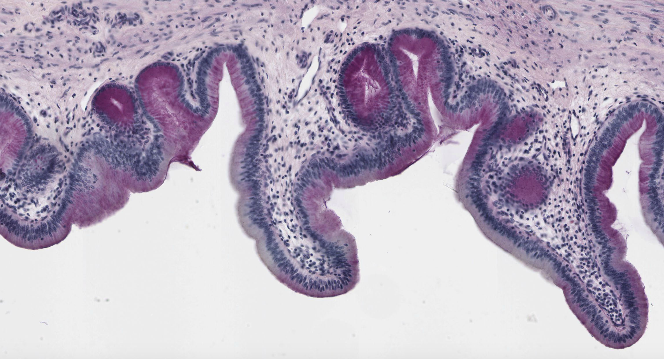
Upon gross examination of slides 194 and 195M (i.e. with the naked eye or at the lowest power on the virtual microscope) you will see a portion of the gall bladder wall nestled in an indentation of the liver tissue. Examine the wall of the gall bladder with your microscope. Extensive folds of the mucosa extend into the lumen. The mucosa consists of a tall, simple columnar epithelium and its underlying connective tissue (constituting a lamina propria). No submucosa is defined. The muscularis consists of scattered bundles of smooth muscle. Deep to the muscularis is an adventitia consisting of rather dense connective tissue that binds the gall bladder to the liver. Where the surface of the gall bladder faces the abdominal cavity there is a serosa.
224 Gall bladder epithelium Gall Bladder Epithelium (Simple Columnar Epithelium) View Virtual EM Slide
Review the role of the gall bladder epithelium in absorption and concentration of bile.
1. Which of the following statements regarding the three zones comprising the liver acinus (of Rappaport) is CORRECT?
- Zone 1 is closest to the central vein.
- Zone 2 is the first to undergo necrosis if circulation is impaired.
- Zone 3 is closest to branches of the hepatic artery.
- Zone 1 is the first to receive nutrients delivered by the portal vein.
- Zone 2 is the last to receive any toxins that may be in the blood.
Answer
Correct answer 4. Zone 1 is the first zone to receive nutrients, which are supplied by the portal vein and are delivered to the sinusoids by the distributing venules.
2. The organ/region of the GI tract is depicted in the blow micrograph?
View Image
- The lower esophagus
- The cardia of stomach
- The pylorus of stomach
- The duodenum
- The gall bladder
- The jejunum
- The appendix
- The colon
Answer
Correct answer 5., a gall bladder.
3. Identify the location of the black asterisk.
View Image
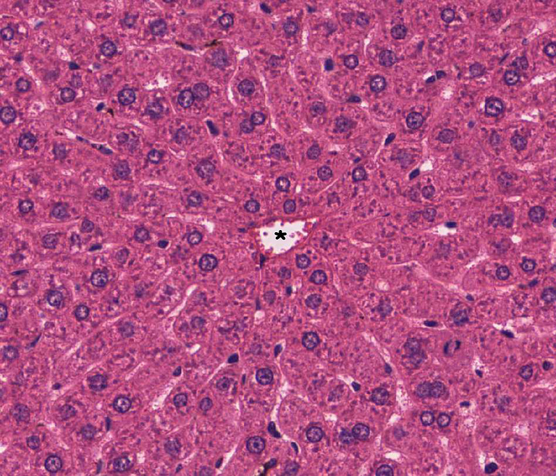
- A liver sinusoid
- The space of Disse
- A central vein
- A branch of hepatic artery
- A bile duct
- A pancreatic intercalated duct
- A pancreatic interlobular duct
Answer
Correct answer 3. Noting the abundance of hepatocytes, the image is from a liver. The vessel is not part of a portal triad, leaving a central vein as the most likely answer.
Pancreas
- Discriminate between exocrine and endocrine pancreas
- Discuss the cellular structure of an exocrine pancreatic acinus and its function
- Identify intercalated ducts and centroacinar cells
- Recognize Islets of Langerhans (endocrine pancreas)
- Discuss the various types of endocrine pancreas cells
Slide 188 pancreas H&E View Virtual Slide
Slide 188B pancreas View Virtual Slide
Tasks:
- Overview of exocrine and endocrine pancreas
- Identify pancreatic acini with centroacinar cells
- Find intercalated duct and interlobular ducts
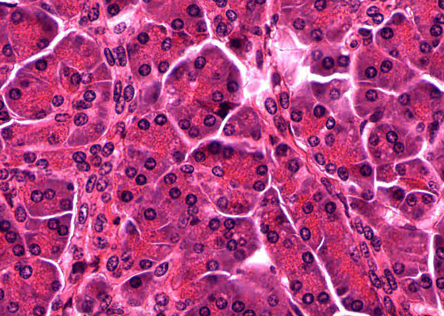
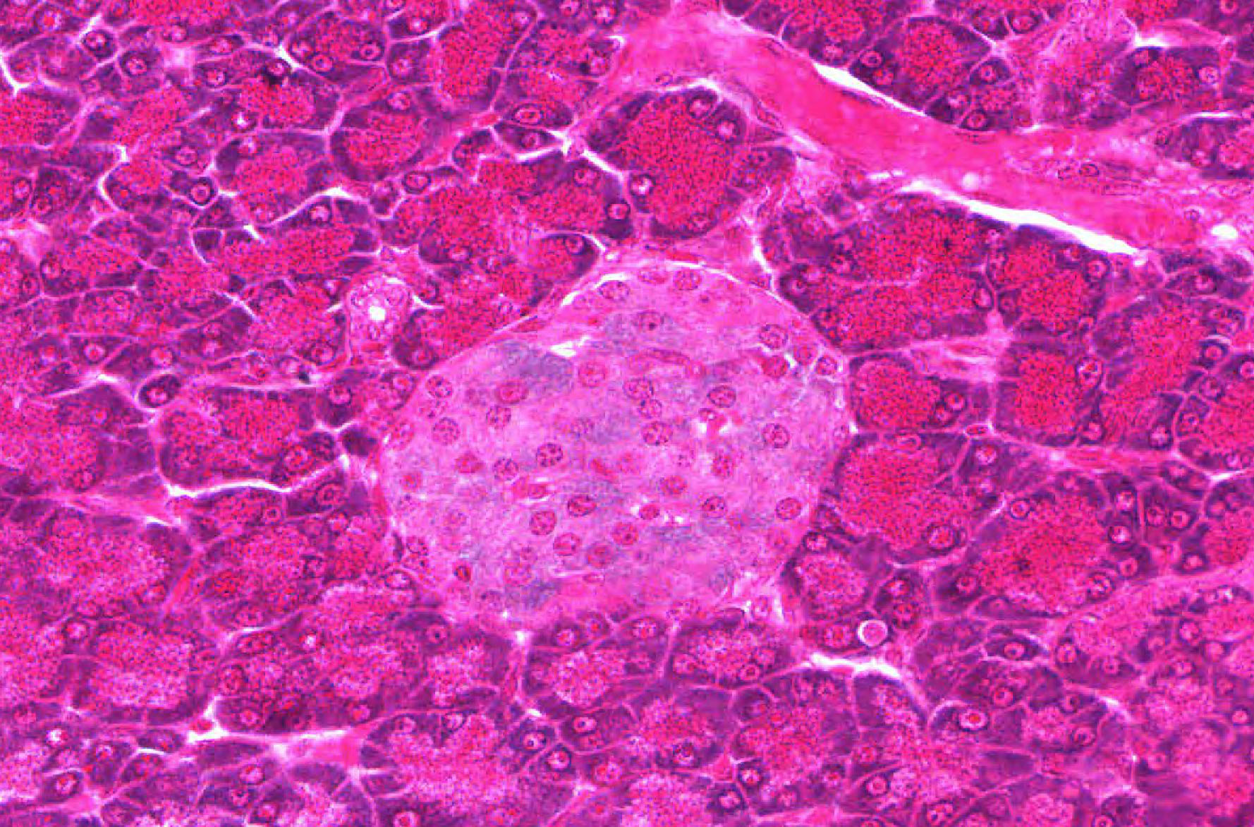
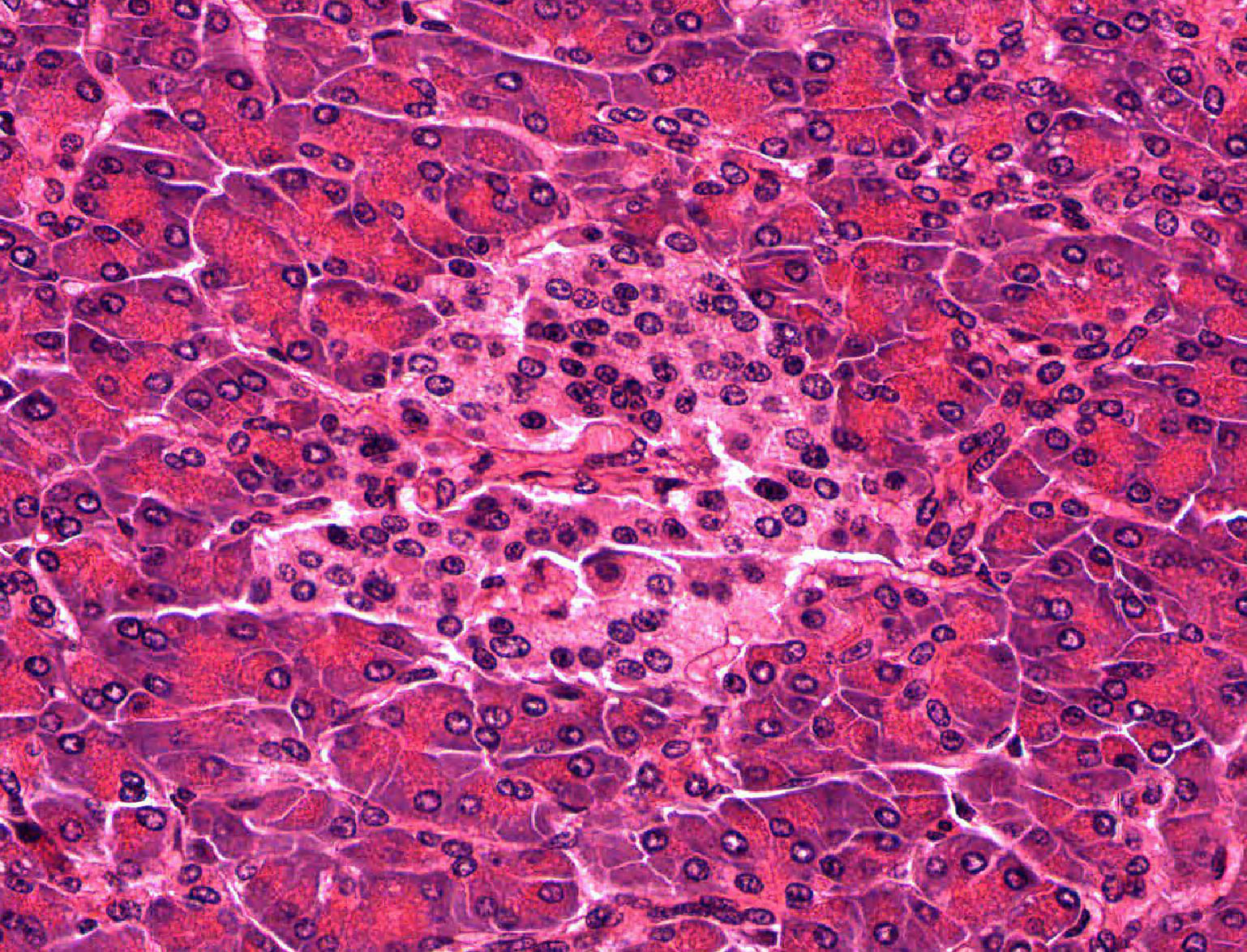
Examine slide 188 at the lowest power and note that most of the section appears purple or bluish. This is the parenchyma (or functional tissue) of the exocrine pancreas. You will note that the parenchyma is rather indistinctly divided into smaller areas by slits of open space or by pink connective tissue (stroma). The smallest of these areas constitute the lobules of this gland. You may see a few circular structures of various size between the lobules. These are cross sections either through branches of the pancreatic duct or through blood vessels. If you observe the parenchyma carefully you will note scattered small spots that are a lighter blue-gray. These are the islets of Langerhans, which comprise the endocrine pancreas.
First observe the parenchyma, noting that it is made up of large numbers of acini , although you may also see occasional fat cells in the parenchyma. Each acinus is a cluster of secretory cells arranged around a small lumen (which is generally collapsed and therefore not visible in your sections). The acini may vary considerably in shape, since they are cut randomly in the section. Note that the peripheral region of each acinus, which represents the basal portions of the individual acinar cells, stains more blue or purple. The hematoxylin component of the H&E stain is staining the ribosomal RNA in the abundant rough (or granular) endoplasmic reticulum found in this portion of the secretory cells.
This “cytoplasmic basophilia” is the reason why the whole section appears purple or blue. The central region of the acinus, representing the apical portions of the acinar cells, is pink (acidophilic) because of the presence of the Golgi complex and numerous secretory granules in this part of the cell (you will probably not be able to make out the individual granules). Here and there you may see a smaller cell, or cluster of cells, with pale cytoplasm in the central region of an acinus. These are centroacinar cells slide 188B View Image and represents the initial portion of the excurrent duct that extends up into the acinus. These slender ducts extending from the acini to larger excretory ducts located outside the lobule are called intercalated ducts slide 188B View Image and may be found by looking for small clusters of 3-5 slightly elongated nuclei lying between the acini; the cytoplasm of the duct cells is very pale, and you may or not be able to make out the lumen. As in salivary glands, intercalated ductal cells in the pancreas contribute bicarbonate ions (sodium and water follow passively) to the exocrine secretory product. However, unlike salivary glands, there are no striated ducts in the pancreas to recover sodium, so the final product is rich in both sodium and bicarbonate (as opposed to saliva in which the sodium content is about one tenth that of plasma).
Using intermediate or low power, observe the larger ducts that are located in the connective tissue septa between the lobules. These interlobular ducts slide 188B View Image can be distinguished from blood vessels by their lining epithelium, which is either simple cuboidal or, in the larger ducts, simple columnar.
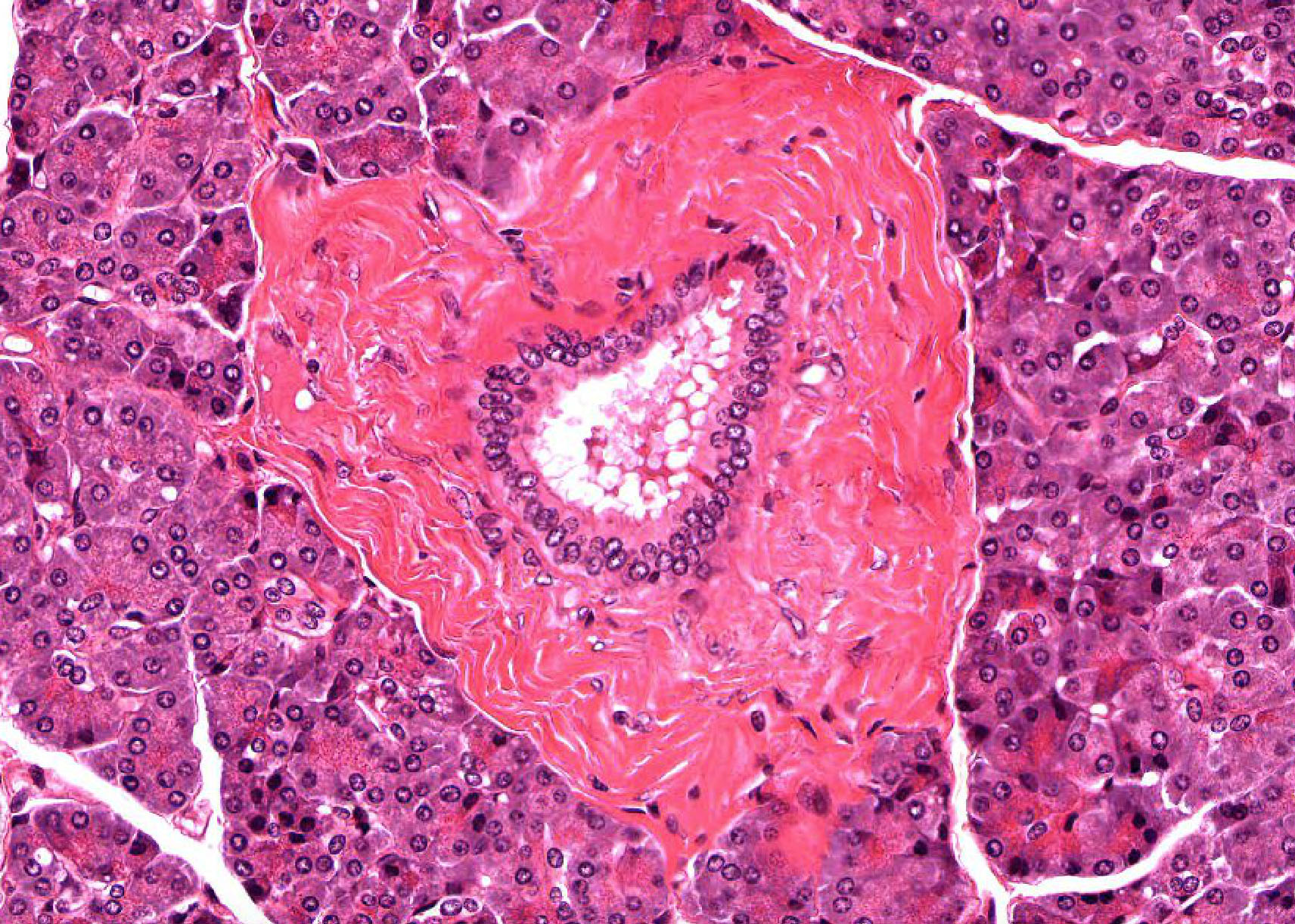
Slide 190B pancreas (rat) chrome-alum hematoxylin and phloxine stain View Virtual Slide
Tasks:
- Identify islets of Langerhans
- Look at Hematoxylin and Phloxine stained pancreatic tissue (Beta cells blue more central, alpha cells more peripheral reddish).
Scan the parenchyma of this slide to find islets. The staining procedure used here allows you to differentiate the two principal cell types found in the islets in slide 190B islet View Image. Although the nuclei in both a and b cells are reddish, the insulin secretory granules in the beta (or B) cells cause the cytoplasm to stain a pale blue-green with the chrome-alum hematoxylin. The alpha (or A) cells, containing secretory granules of glucagon, are stained reddish. Note that the beta cells are usually more numerous and occur in the interior of the islet, while the alpha cells are found more peripherally. You will not be able to distinguish delta (or D) cells, source of somatostatin. Incidentally, the secretory granules of the acinar cells are seen clearly in the exocrine pancreas in this slide. Islets of Langerhans can also be readily seen in slide 188B Islets of Langerhans View Image as well. The islets occur as pale areas of cells here and there in the parenchyma (you can find them most easily under low power). Note the scattered distribution of the islets and their variation in size. You will not be able to distinguish the various cell types in the islets in this routine H&E preparation.
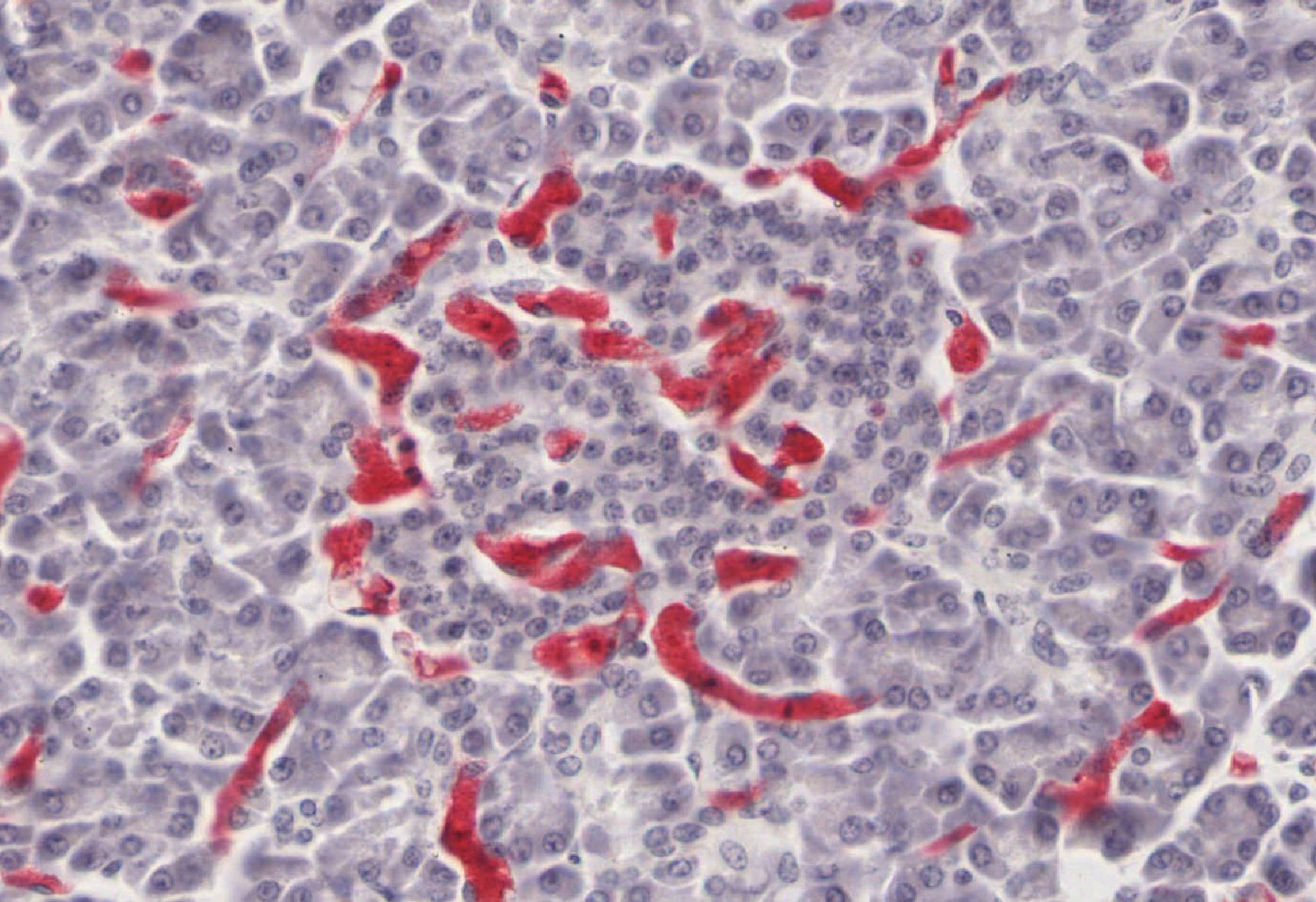
Slide 189 Pancreas (monkey) vascular injection TB&E View Virtual Slide
Task:
- Look at the vascular perfusion of islets of Langerhans.
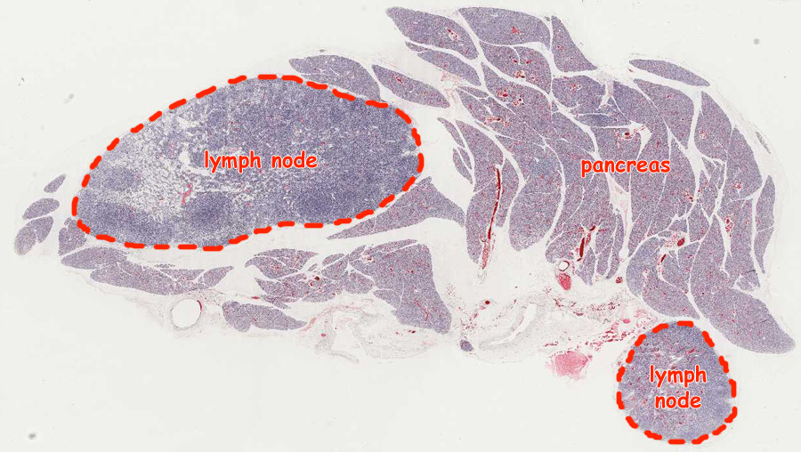
Before histological preparation, the arterial supply of this pancreas was injected with a red material. The main point of the slide is to show you how much richer the vascular supply is to the endocrine tissue, the islets of Langerhans, than to the surrounding exocrine pancreas. Find islets in the parenchyma, and observe the denser concentration of capillaries slide 189 dense capillary concentration View Image. Many of these slides in our collection (including the virtual slide) contain some pieces of lymph nodes, so make sure that you are looking at the pancreatic tissue orientation Image
226 Pancreas Exocrine Pancreas View Virtual EM Slide
In this low power electron micrograph, observe the organization of the acini, composed of acinar cells. Within the acinar cells you will see the basal rough endoplasmic reticulum and the numerous secretory granules in the apical region of the cells, facing the small lumen of the acinus. Note the centroacinar cell in upper right acinus.
227 Pancreas - Exocrine, detail of acinus Organelles of the Secretory Pathway View Virtual EM Slide
Pancreatic acinar cells as depicted in this electron micrograph are cells that are highly specialized for protein secretion. Therefore, all the organelles of the protein secretory pathway are well-represented and are clearly visible in this micrograph.
230 Pancreas Endocrine Pancreas View Virtual EM Slide
You will not be ask to identify different types of endocrine cells in the islet of Langerhans. However, compare the appearance of an endocrine cell containing small granules to that of a portion of exocrine cell shown on the right.
1. Which statement about the cell indicated by the black arrow is true?
View Medium Mag Image
View High Mag Image

- It produces bile.
- It is located in the space of Disse.
- It produces pancreatic pro-enzymes (such as trypsinogen).
- It adds bicarbonate and water to the pancreatic exocrine secretion.
- It removes sodium from the pancreatic exocrine secretion.
- It secretes insulin.
- It secretes glucagon
Answer
Correct answer 4. The arrow indicates an intralobular or intercalated pancreatic duct cell, which adds bicarbonate and water to the pancreatic exocrine secretion.