From the lecture
- Understand the processes of preparing and viewing tissues by light and electron microscopy.
- Understand the physical bases for the appearance of tissues in the light and electron microscopes (e.g. What is basophilia and what causes structures to be basophilic? What creates the contrasting light and dark regions in an electron micrograph?)
From the lab session
- A brief listing of some common stains is present at the end of this section. You should have a general familiarity with H&E (Hematoxylin and Eosin), Masson, PAS, and elastic stains.
- Become familiar with the various ways to access and view images in the Michigan virtual slide collection
Hematoxylin and Eosin Stain (or H&E Stain)
Hematoxylin is the most commonly used nuclear stain in histology and pathology although, despite its long use and honorable history, the chemistry of the dye is still not fully understood. Essentially, hematoxylin is a basic dye and complexes with nucleic acids (DNA and RNA in the nucleus; RNA in the cytoplasm) or other negatively charged molecules (such as sulfate groups). Structures that bind hematoxylin are therefore termed "basophilic" (base loving).
Eosin is an acidic dye and the basic structures it stains are termed "eosinophilic" or less commonly "acidophilic" (acid loving). It stains membranes and most proteins. Cells that have large quantities of folded membranes stain intensely with eosin, because of basic amino acids in the membranes (e.g. macrophages contain lots of membrane in the form of phagocytic vesicles as well as basic lysosomal enzymes within those vesicles that stain with eosin). Collagen is generally stained some shade of red/orange whereas actin (such as in smooth muscle cells) is a bit more pink. Elastin, when present in relatively large amounts (such in the walls of blood vessels, in elastic cartilage, and in the esophagus and trachea), will appear glassy red.
A note about acids/bases and their charges: It always seems to a point of confusion as to how it is that an acid such as DNA can have a negative charge when we generally think of something that is acidic as being positively charged (i.e. a solution with lots of H+ ions is "acidic"). However, the better way to think of acids is as proton donors --in solution, an acid such as DNA donates H+ protons (which makes the solution acidic). Upon donating protons, the DNA therefore becomes negatively charged and it is in this state that it binds hematoxylin.
Good examples of H&E-stained sections in the Michigan Histology collection:
Slide 029 (small intestine) View Virtual Slide
Slide 106 (thick skin) View Virtual Slide
Masson Triple or Trichrome Stain
This dye combination stains mucus as well as collagenous and reticular fibers blue (aniline blue) or green (fast green) depending on the mixes of dyes used; muscle red; nuclei red (they are black if preceded by an iron hematoxylin). This is a commonly used connective tissue stain in both histology and pathology. On your slides the stain is designated "Masson" or "Mass"; but the blue or green collagen is the tip-off. This staining method was developed by the Canadian physician Claude L. Pierre Masson (1880-1959).
Reference:
Masson, P.: Some histological methods. Trichrome stainings and their preliminary technique. J. Tech Methods 12: 75-90 (1929)
Good examples of Masson Trichrome-stained sections in the Michigan Histology collection:
Slide 098N (heart) View Virtual Slide
Slide 152AF (oral pharynx) View Virtual Slide
Elastic Fiber Stains
- Aldehyde Fuchsin Stain also known as Gomori's aldehyde-fuchsin stain after the Hungarian-American physician and histochemist George Gömöri (1904-1957)
- Aldehyde fuchsin is a deep purple dye. It stains elastic fibers and granules of beta cells in the islets of Langerhans, cartilage matrix, and stored neurosecretory product in the hypophyseal pars nervosa, among other things. In some of the Michigan collection slides, it is the only stain and therefore only elastin is demonstrated. For other slides it is combined with Masson's trichrome (see slide 044).
Reference:
Gomori, G.: Aldehyde-fuchsin: a new stain for elastic tissue. Amer. J. Clin. Pathol. 20, 665–666 (1950)
Good examples of aldehyde fuchsin-stained sections in the Michigan Histology collection:
Slide 036 (aorta) View Virtual Slide
Slide 044 (ear pinna) View Virtual Slide
- Weigert's Stain or Weigert’s aldehyde stain after the German Jewish pathologist Carl Weigert (1845-1904)
- Uses a different kind of fuchsin (basic fuchsin), but the result is similar: elastic fibers stain a deep purple color.
Reference:
Weigert, C.: Über eine Methode zur Färbung elastischer Fasern. Zbl. Allg. Path. Anat. 9, 289-292 (1898)
The only example of a Weigert's aldehyde-stained section in the Michigan Histology collection is
Slide 100W (heart wall) View Virtual Slide
- Verhoeff or Verhoeff-van Gieson Elastic Tissue Stain named after the American ophthalmologist Frederick Herman Verhoeff (1874–1968) and the American neurologist Ira Van Gieson (1866-1913)
- Verhoeff's hematoxylin contains ferric chloride and iodide which causes it to stain elastic fibers deep purple/black. Frequently counterstained van Gieson's solution with which stains collagen red/orange and cytoskeletal elements (such as actin) yellow-brown.
References:
Verhoeff, F.H.: Some new staining methods of wide applicability. Including a rapid differential stain for elastic tissue. JAMA 11: 876-877 (1908)
Gieson, V.: Laboratory notes of technical methods for the nervous system. New York Med. J. 50: 57-60 (1889)
Good examples for Verhoeff/Van Giessson-stained sections in the Michigan Histology collection:
Slide 033 (skin) View Virtual Slide
Slide 303 (artery and vein) View Virtual Slide
Silver Stain
There are several different types of histological silver stains. Usually silver nitrate is reduced to metallic (black) silver. The process of development and fixation is similar to developing a photograph (stains reticular fibers). Most of the silver-stained slide in the Michigan collection are specific for reticular (collagen III) fibers. Slide 198-1 View Virtual Slide is different as it stains the bile canaliculi network in liver tissue.
Reference:
Puchtler, H. and Waldrop, F.S. Silver impregnation methods for reticulum fibers and reticulin: A re-investigation of their origins and specifity. Histochemistry 57, 177–87 (1978)
Good examples of silver-stained sections in the Michigan Histology collection:
Slide 28-2 (lymph node) reticular fiber staining View Virtual Slide
Slide 198 (liver) staining reticular fibers in the space of Disse View Virtual Slide
Periodic Acid Schiff (PAS) Stain
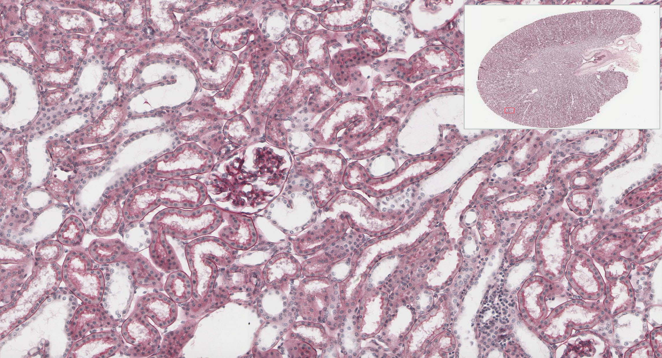
This is an extremely useful technique for demonstrating glycoproteins, mucins and some proteoglycans -anything that contains a relatively high amount of sugar groups. It involves the generation of dialdehydes from hexoses (present as the carbohydrate portion of the aforementioned compounds and is based on the chemistry (Schiff bases) developed by the German-Italian chemists Hugo Schiff (1834-1915). One of its main uses is the demonstration of basement membranes, especially in the kidney, and/or in sections with epithelia atypia, where breech of the basement membrane is suspected in early carcinomas. An excellent example is slide 210 View Image of a kidney where PAS staining demonstrates the basement membranes (pink lines) of the simple cuboidal epithelium lining the tubules and squamous epithelium in the glomeruli (the round tangles of cells). Note that PAS staining also shows the glycocalyx associated with microvilli (appears as a fuzzy pink border) on epithelia lining some of the tubules.
References:
Schiff, H.: Mittheilungen aus dem Universitätslaboratorium in Pisa: Eine neue Reihe organischer Basen. Justus Liebigs Ann. Chem. 131, 118-119 (1864)
Yamabayashi, S.: Periodic acid—Schiff—Alcian Blue: A method for the differential staining of glycoproteins. Histochem. J. 19, 565-571 (1987)
Good examples for PAS-stained sections in the Michigan Histology collection:
Slide 160 (gastro-esophageal junction) with Azure Blue counterstain View Virtual Slide
Slide 210-PAS (kidney) with hematoxylin counterstain View Virtual Slide