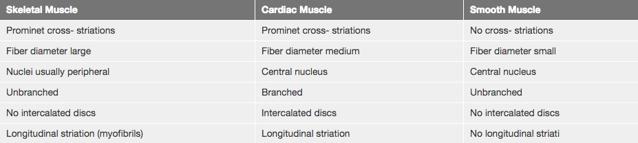
- Name and identify the three types of muscle at the light and EM level and name a few distinctive features for each.
- Describe and understand the structural basis of muscle striation at the molecular, as well as at the light and EM microscope level and how different structures change during muscle contraction.
- Know the structural elements and the general molecular mechanisms that underlies muscle contraction for all three muscle types (skeletal, cardiac and smooth muscle).
- Be familiar with the regenerative potential of each muscle type.
There are three major types of muscle, and their structure reflects their function. Skeletal and cardiac muscle cells are called striated muscle because of the very regular arrangement of their intracellular contractile units, sarcomeres, at the light microscope (LM) and electron microscope (EM) levels. This regular arrangement imparts a cross-striated (or striped) appearance. Such an arrangement is not seen in smooth muscle cells. Skeletal muscle is also called voluntary muscle, because its contraction is under conscious neural control. In contrast, cardiac and smooth muscles are called involuntary muscles because their contractions are either spontaneously generated or are under the control of the autonomic nervous system.
Each of these three types of muscle has a characteristic appearance in both cross and longitudinal sections. You should be able to recognize each type of muscle in both planes of section.
A. Cytology of skeletal muscle cells
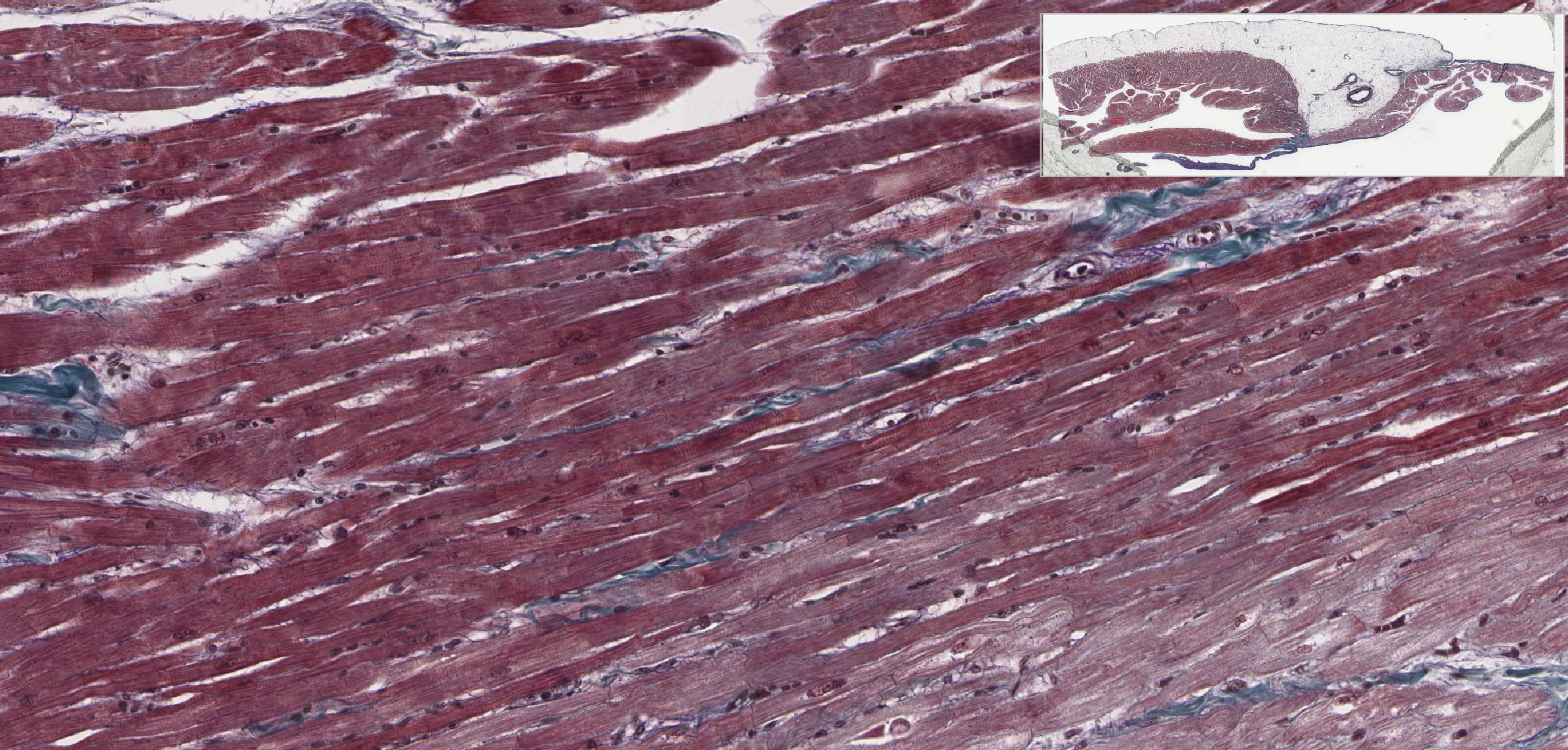
Slide 058L skeletal muscle H&E longitudinal View Virtual Slide
Slide 058Lex Skeletal muscle H&E longitudinal View Virtual Slide
Slide 058Thin skeletal muscle H&E longitudinal View Virtual Slide
Slide 058T Skeletal muscle H&E cross View Virtual Slide
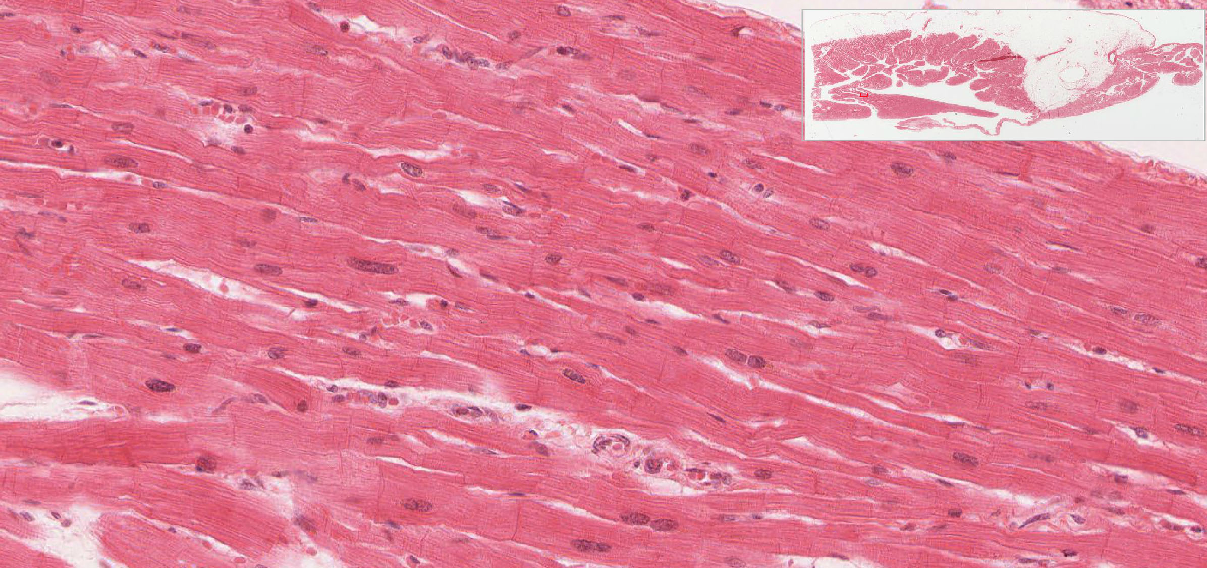
In longitudinal sections of skeletal muscle (Slide 58), observe the following :
- Nonbranching, cylindrical shape of the cell (also referred as muscle fiber). These cells are very long; you cannot see their ends.
- Be sure you can identify the borders of the muscle cell. You might see occasional nuclei which appear to be centrally located, but aren't. Why?
Answer
Again the answer to the question is in the plane of section. Cells with peripheral nuclei that are cut at an angle can appear to have centrally located nuclei. This is why it is important to look at other features of the cell to determine what exactly it is.
- Peripheral position of the elongate nuclei just inside of the sarcolemma (plasma membrane). Note that each cell contains large numbers of nuclei.
- Cross striations can be seen, and are due to the structure of the sarcomere. A sarcomere consists of the structures between two Z lines. You should recognize:
- The dark A band
- The lighter H zone which bisects the A band
- The light I band d. The dark Z line which bisects the I band.
It is admittedly difficult to see the Z line and especially the H zone with the light microscope. However, they can be seen clearly on some areas of almost all slides, and it is just necessary to do some looking around for a favorable area on your slide. Some good areas of skeletal muscle showing cross striations can be found in slide 058thin View Image.
- A less regular longitudinal striping can sometimes be seen within the muscle fibers and is due to the bundling together of the thick and thin filaments into myofibrils, which are arranged parallel to the long axis of the cell. Note that sometimes the cross striations are not aligned all the way across the cell. This is because different myofibrils may not be aligned.
In transverse sections of skeletal muscle cells (slide 58), observe the cylindrical shape of the cells (fibers) and the peripherally-located nuclei. Note also the the cytoplasm of the muscle cells has a stippled, punctate appearance which is due to the bundling of thick and thin filaments into myofibrils as mentioned above. Know what exactly is a muscle cell and what's the difference between a muscle fiber and a muscle fibril. The basic units of muscle are the contractile proteins actin and myosin arranged in sarcomeres. Placed end to end, these sarcomeres form long bands called myofibrils. Within a skeletal muscle cell, the numerous myofibrils are separated by glycogen, mitochondria, and muscle triads (two terminal cisternae and a T tubule) and other organelles. Chances are, if you are looking at an electron micrograph of muscle tissue, you are looking at myofibrils. Groups of these fibrils form a muscle fiber, which is surrounded by endomysium. Muscle fiber and muscle cell are synonymous. Groups of individual cells that are surrounded by perimysium are known as fascicles and groups of these fascicles, surrounded by epimysium make up a muscle.
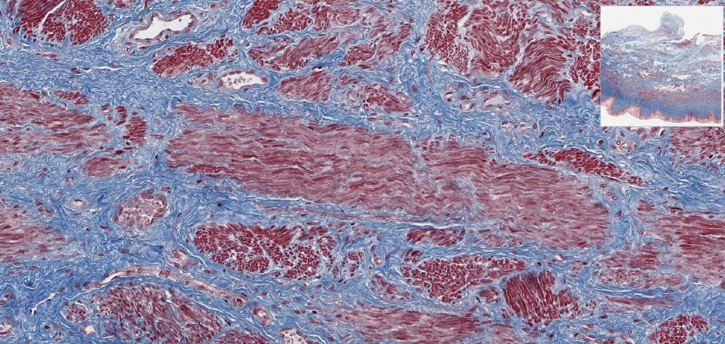
B. Perimuscular Connective Tissue
Slide 059-3 forearm muscle Masson cross fetal View Virtual Slide
Slide 059-1 forearm muscle H&E cross fetal View Virtual Slide
In slide 59, stained with trichrome or H&E, three layers of connective tissue sheaths are visible. This is a transverse section of an entire fetal forearm, and contains many elements in addition to the muscle, such as nerves and tendons, both of which are, unfortunately, simlarly bundled into fascicles and therefore easily confused with muscle. So, make sure you're looking at the muscles
- Endomysium - thin connective tissue sheath, consisting of basal lamina, some reticular fibers and capillaries around each muscle fiber. In fetal muscle, endomysium is not always clearly defined, so it is best to use the trichrome-stained slide (slide 59-3) to this - it is the connective tissue immediately surrounding the individual cells and very delicate perhaps visible only as a bluish tinge.
- Perimysium - loose connective tissue sheath consisting of type I collagen fibers found around fascicles (bundles of muscle fibers). Note that the tissue in these specimens underwent some shrinkage during preparation so the separation between fascicles is somewhat exaggerated. Similarly, much of the perimysium has been damaged; however there are still some areas where it can be clearly seen (e.g. around many of the blood vessels found within the belly of the muscles).
- Epimysium - the dense irregular connective tissue sheath around the entire muscle = deep fascia in gross anatomy; contains even larger blood vessels and nerves.
Three layers can be seen in slide 059-3 View Image [see orientation]
![[see orientation]](/sites/default/files/images/slides/2muscle.jpg)
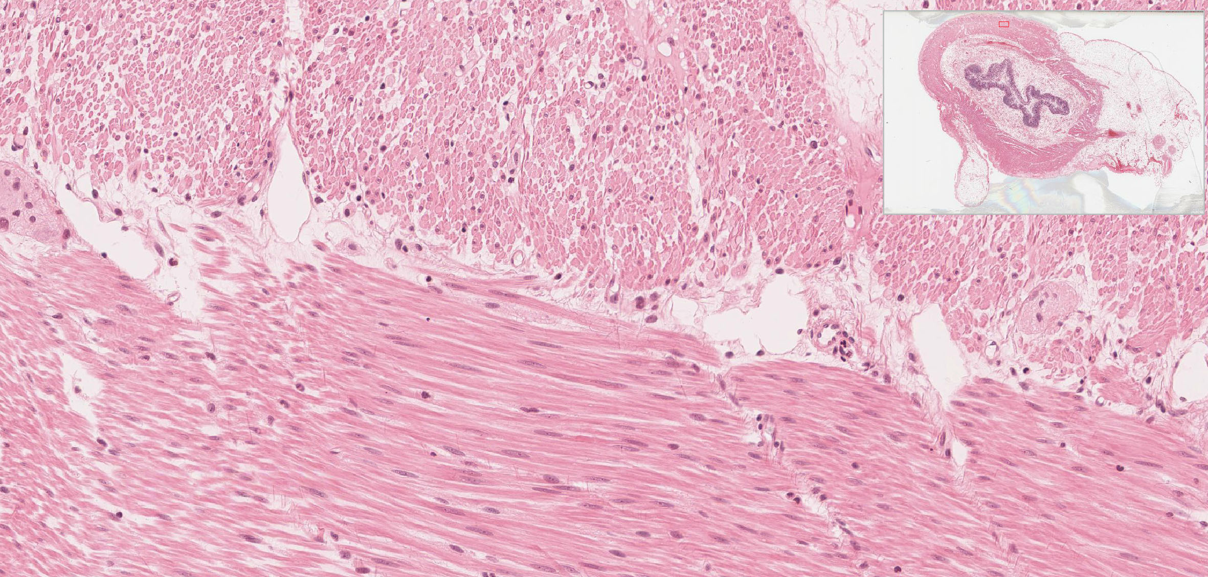
Slide 057 ventricle H&E View Virtual Slide
Slide 098-1 Heart ventricle H&E View Virtual Slide
Slide 098N right wall Masson View Virtual Slide
Slide 305 heart ventricle H&E View Virtual Slide
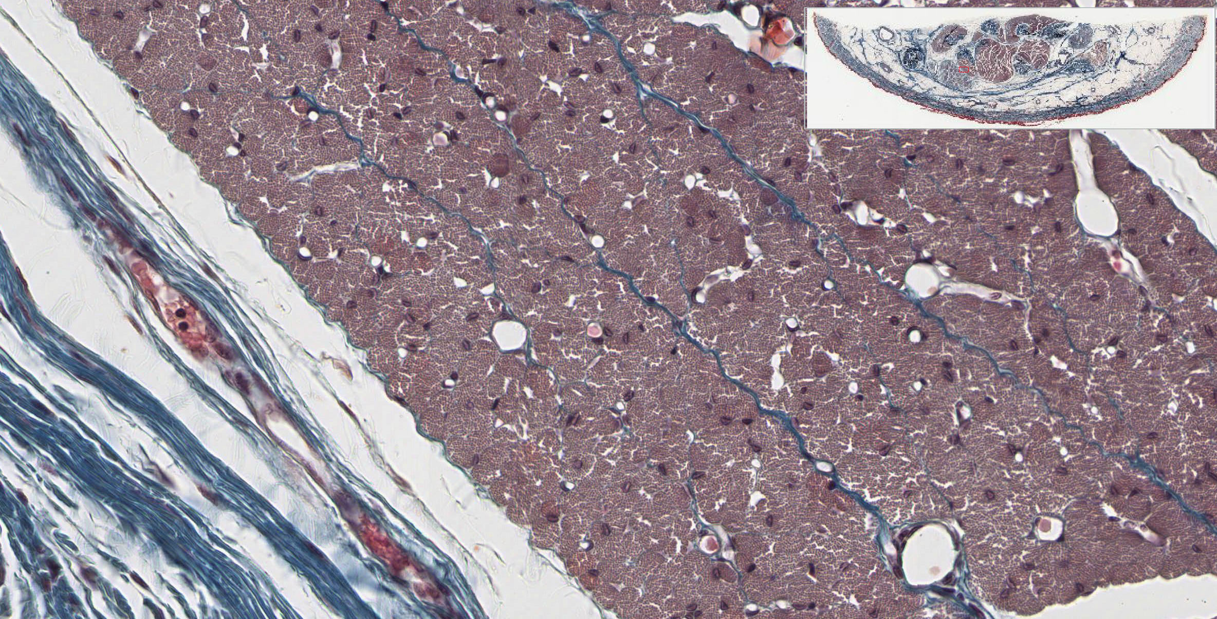
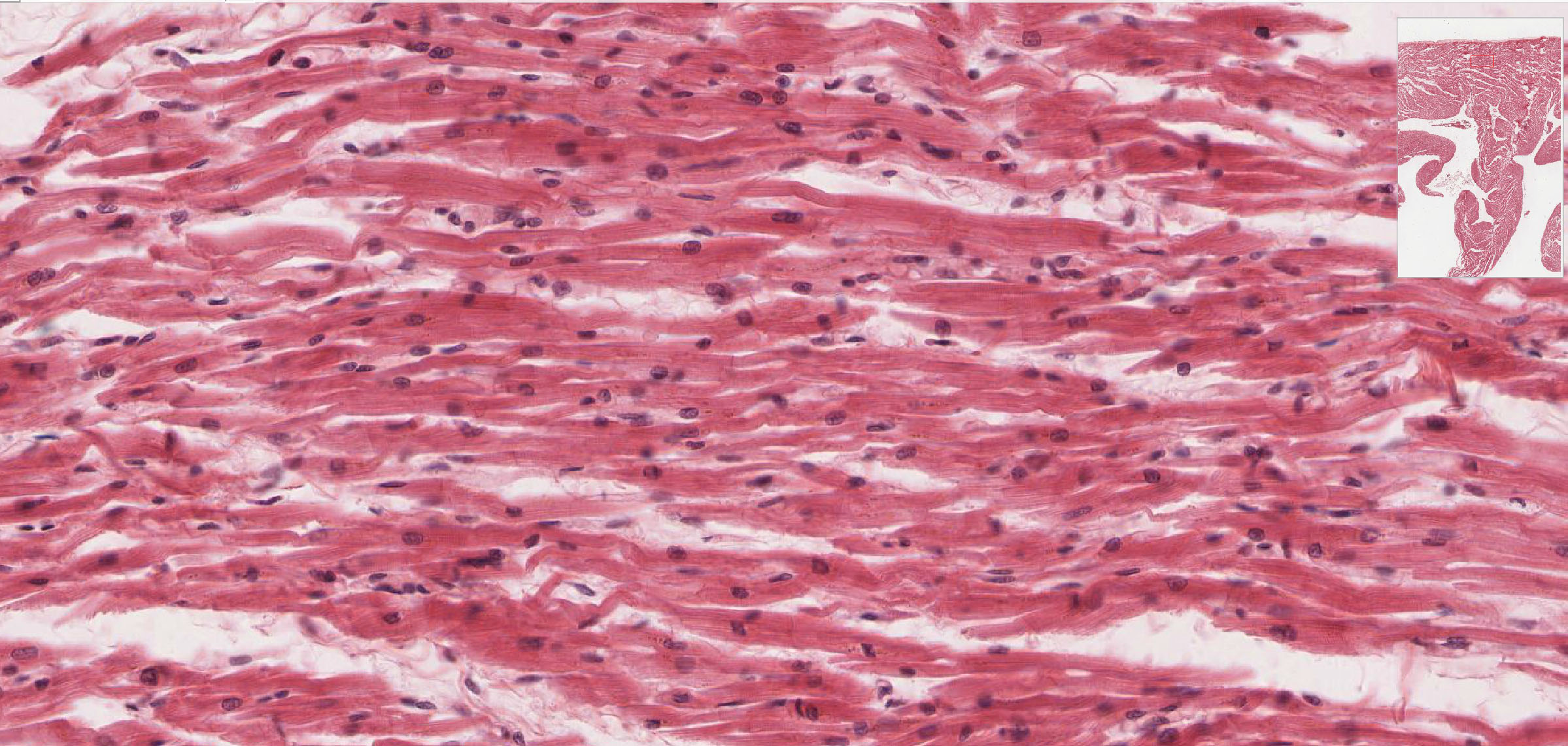
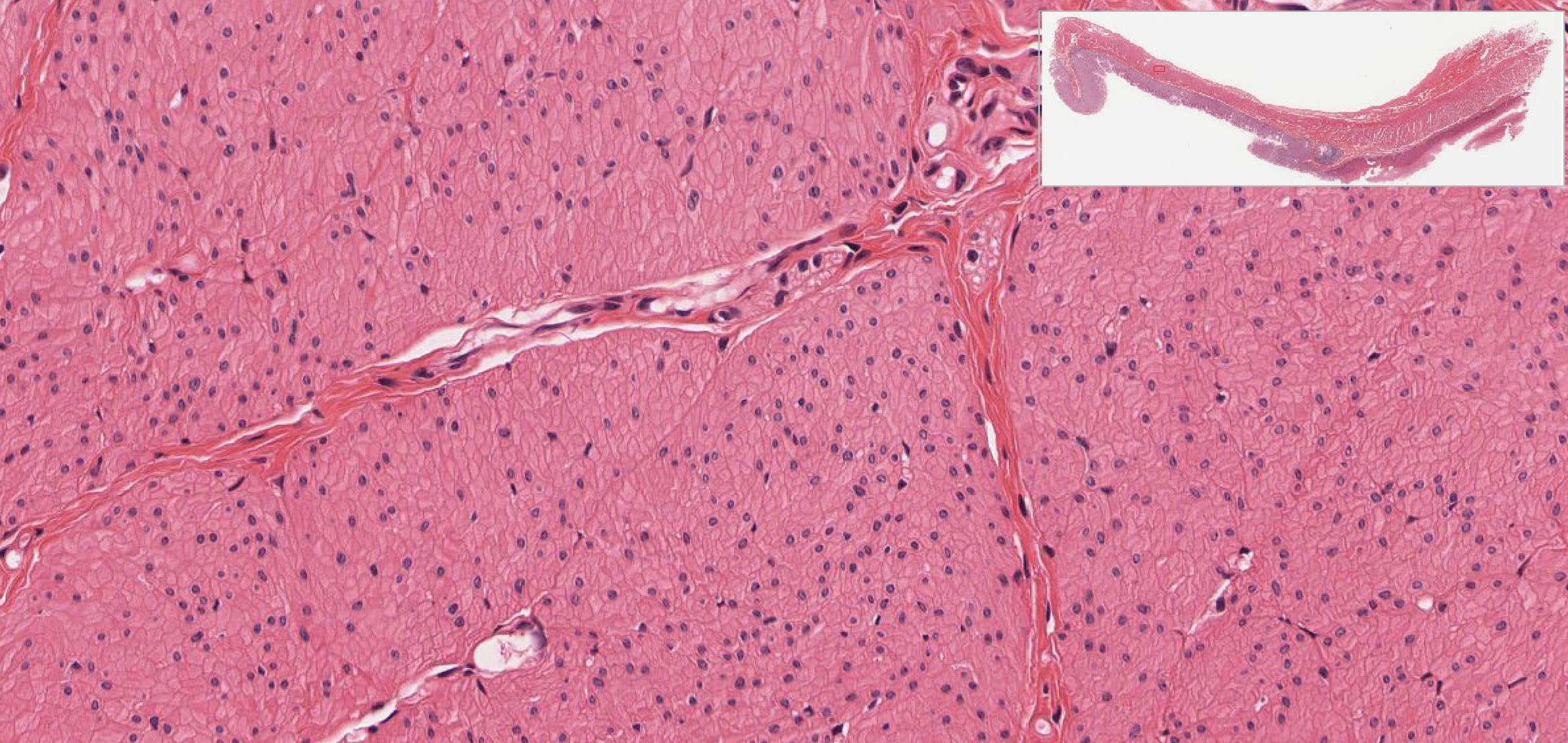
Cardiac muscle will be studied in the wall of the ventricle of the heart. In comparison with skeletal muscle, note the following differences. Cardiac muscle cells branch and form a three-dimensional network. These branch points can sometimes be seen in your sections, and you should also note that the muscle fibers are less parallel than in skeletal muscle.
A. In longitudinal sections, observe:
- Intercalated discs which are dark lines which cross the cell transversely. Look around in slide 057 View Image or slide 305 for lightly stained regions of the slide. There are artifactual transverse breaks in some of the muscle fibers, which are NOT intercalated discs. Intercalated discs are also fairly easy to find in the areas where muscle fibers are longitudinally oriented in slide 098-1 View Image and/or slide 098N View Image (be sure to look at both H&E and trichrome-stained sections).
- The nucleus is centrally located in a fiber.
- Longitudinal striping due to myofibrils can sometimes be seen.
B. In transverse sections, observe:
- The myofibrils are coarse and give rise to the nonhomogeneous, punctate appearance of the sarcoplasm.
- The nuclei are centrally-located. You won't see a nucleus in every fiber cross-section.
- The extensive network of capillaries in the endomysium - heart muscle is ALWAYS beating and therefore always in demand of oxygen and nutrients delivered via the blood.
Make sure you can differentiate between cardiac and skeletal muscle in both longitudinal and transverse sections!
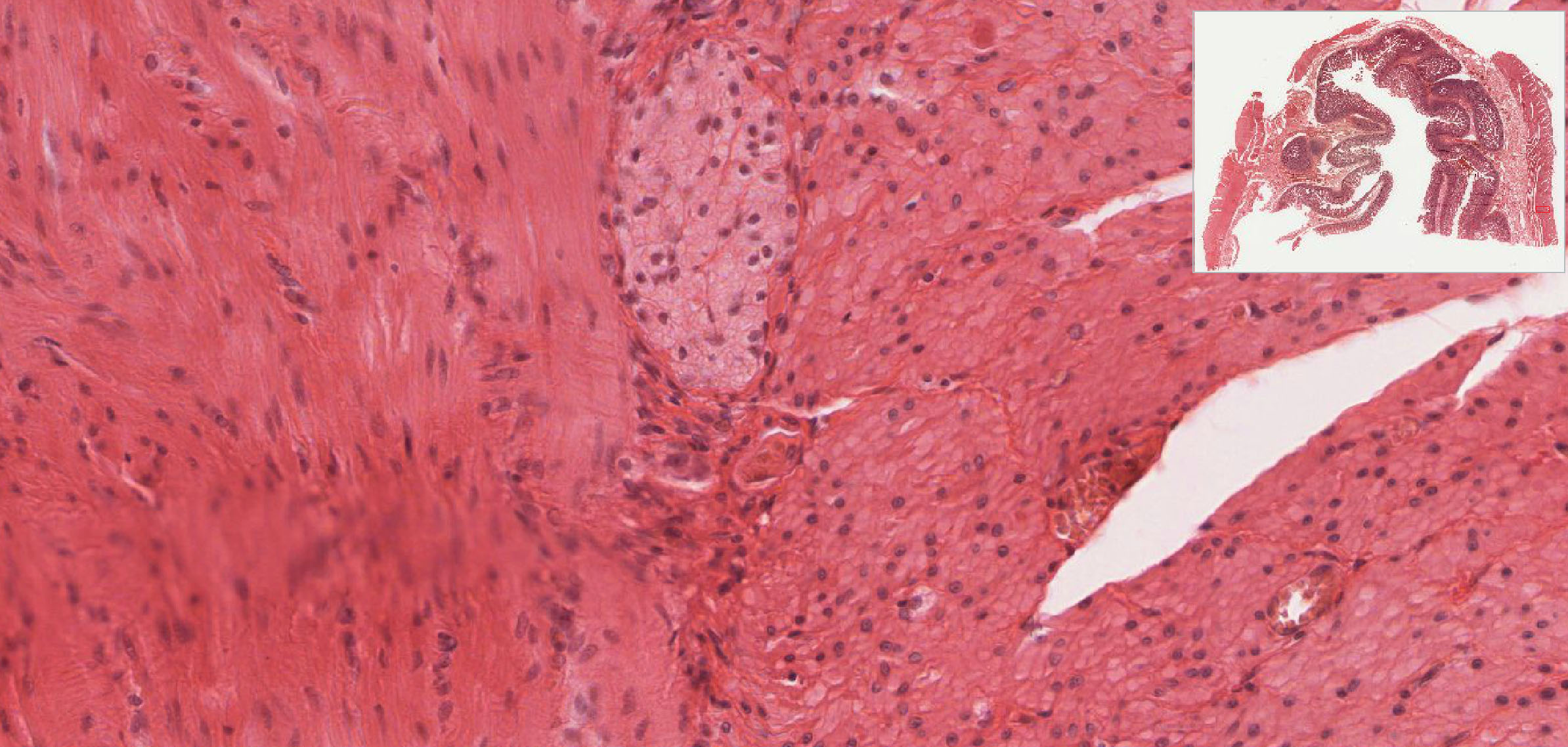
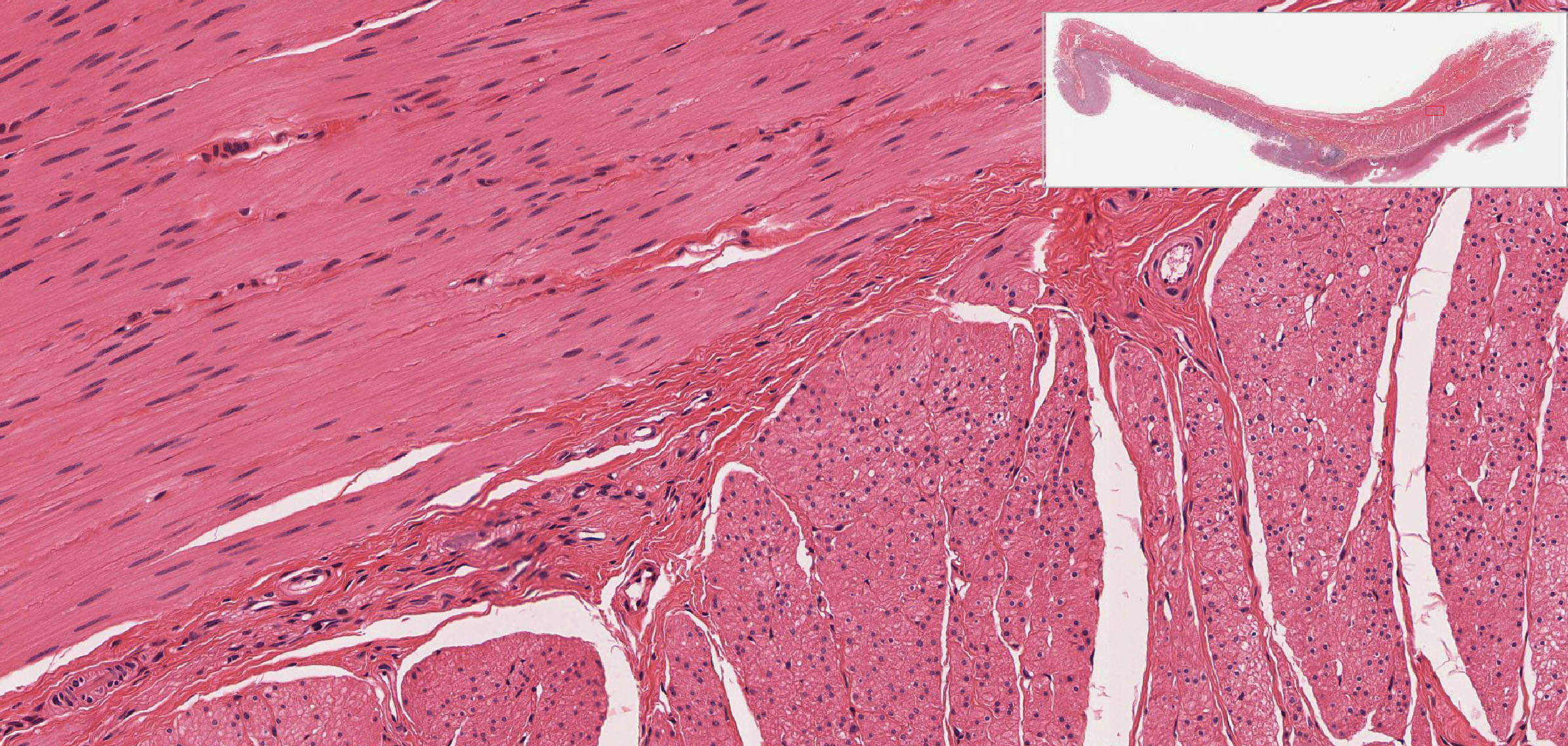
Slide 029-1 Small Intestine (simple columnar epithelium, simple squamous epithelium) H&E cross View Virtual Slide
Slide 169 jejunum H&E cross View Virtual Slide
Slide 155 gastro-esophageal junction H&E longitudinal View Virtual Slide
Slide 250-2 vagina Masson View Virtual Slide
Slide 250-1 vagina H&E View Virtual Slide
Smooth muscle may be studied using slide 029-1 smooth muscle View Image or slide 169 View Image, both in the intestine. To find the muscle layer, look at the at slide at the lowest power (this is about the same as looking at the glass slide with the naked eye). The purple layer is largely the epithelium and the lamina propria filled with plasma cell and lymphocytes. Next to that you see a lighter region of connective tissue (the submucosa you looked at to see loose connective tissue and fibroblasts), then a darker pink region which is made up of the two layers of smooth muscle you want to look at. Slide 29 is a cross section of the intestine, so the inner, circular layer of muscle will have cells oriented longitudinally (or, in places, the cells may appear to be oriented more obliquely). Move further out to see the outer sheet of smooth muscle, which runs longitudinally along the intestine, and will therefore be seen in cross section.
Look at slide 155, which is a longitudinal section of the GI tract at the gastro-esophageal junction, to see more smooth muscle in various planes of section. The smooth muscle in the esophagus (the part lined with a stratified, non-keratinizing squamous epithelium) sample region from slide 155 View Image is organized in the "classic" inner circular and outer longitudinal arrangement (top half in longitudinal section and bottom half in cross section). However, the stomach (the part lined by a columnar epithelium) has an inner oblique layer, a very prominent middle circular layer sample region from slide 155 View Image (seen mostly in cross section here), and a sometimes less obvious outer longitudinal layer. Don't worry knowing about the specific layers or being able to tell esophagus from stomach. However, you should definitely be able to identify smooth muscle in any plane of section (tranverse, longitudinal, or even oblique). In this particular slide, both the hematoxylin and eosin staining are quite intense, which should help you to see the cytoplasm more clearly, especially when the muscle is cut in cross section.
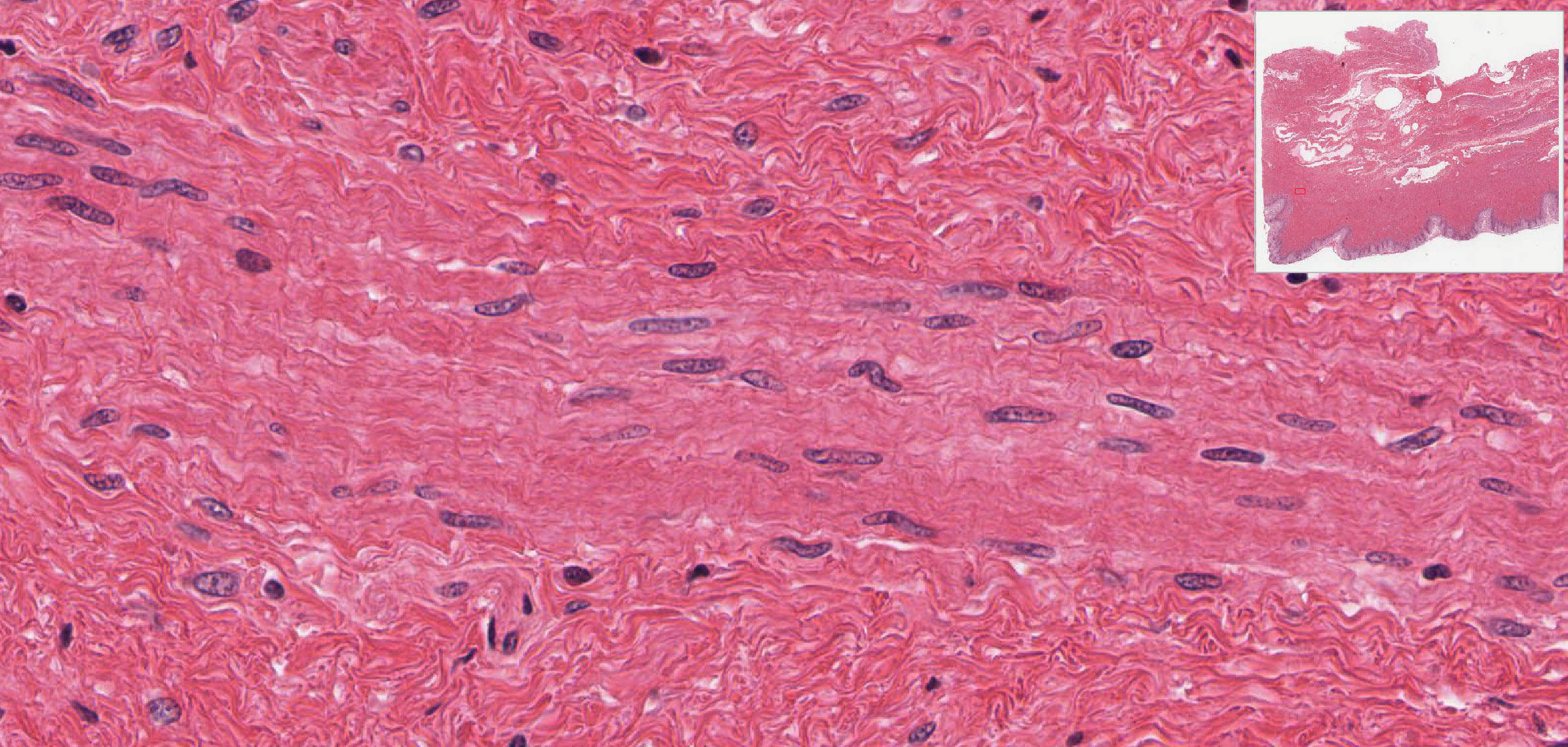
A. In longitudinally cut smooth muscle cells, observe the following points:
- Cells are small and spindle-shaped (fusiform); this may be hard to appreciate because the cell membrane is indistinct.
- Myofibirils and cross striation cannot be seen.
- Nuclei are narrow, elongated and sometimes kinked or spiraling. They are centrally located.
B. In transversely cut smooth muscle cells, observe the following points:
- The cell has a small diameter.
- The nucleus is located centrally, but will not be seen in every cross section.
- Myofibrils cannot be seen.
- Cross-sectional diameters vary due to the spindle shape of the cells.
Now, look at slide 250 and see if you can distinguish between small fascicles of smooth muscle and collagen fibers in the lamina propria (this task will be easier if you look first at the trichrome-stained section, which stains the muscle pink(ish) and the collagen blue) slide 250-2 View Image. It's more challenging to make this distinction in the H&E-stained section slide 250-1 View Image. You should note that smooth muscle is pink, wheras collagen is a bit more orange-red. Also, smooth muscle tissue is mostly cellular (and therefore more nuclei are present), whereas the connective tissue is mostly extracellular collagen fibers with fewer cells. The table below compares the differences in the morphology of the three types of muscle.
Major Histological Characteristics Of The Three Types Of Muscle As Seen With The Light Microscope
37 Skeletal Muscle - Longitudinal Section low Magnification View Virtual EM Slide
Skeletal Muscle (longitudinal section, low magnification). Find the skeletal muscle nuclei and note their peripheral location. Note the intimate contact between capillaries and muscle cells and be sure you can tell where one muscle cell or fiber stops and another begins (you can see parts of four fibers in this picture). Make sure you know which is the longitudinal axis of the cell. Identify sarcomeres, A bands, I bands, Z lines and H zones. Note that, as you saw at the LM level, the individual myofibrils do not line up perfectly across the fiber.
34 Skeletal muscle - Cross Section Low Magnification View Virtual EM Slide
Skeletal Muscle (cross section, low magnification). Note location of muscle fiber nuclei. You can see cross sections of A bands (darker) and I bands (lighter) side by side in the same cell because of the fact that the myofibrils don't line up perfectly. Identify the approximate outline of a single myofibril.
32 Striated muscle - longitudinal section View Virtual EM Slide
Skeletal Muscle (longitudinal section). Identify a sarcomere. Relate the sarcomeric structure seen in the LM to the structure seen here. Note that there is also lots of glycogen in the region between the two myofibrils in this picture, a storage form for glucose (which is metabolized to provide energy for muscle contraction). At the border of the I and A-bands, note triads consisting of a central T (transverse) tubule and flanking cisternae of the sarcoplasmic reticulum.
41 Cardiac muscle Intercalated Disc View Virtual EM Slide
Cardiac Muscle (Intercalated Disc, longitudinal section). Note the somewhat irregular course of the intercalated disc. In this preparation, the I bands are very short, indicating that the sarcomere is in a contracted state. Review the types of junctions present in an intercalated disc and their functions.
43 Cardiac Muscle View Virtual EM Slide
Cardiac Muscle (longitudinal section). Note central location of muscle nuclei. Note the "stacks" of mitochondria between myofibrils. Cardiac muscle is even richer than skeletal muscle in mitochondria (again, important for energy production). An intercalated disc is present in the upper left region of the picture.
207 Small intestine (Muscularis Externa) View Virtual EM Slide
Study the orientation of the smooth muscle cells in the intestinal muscularis externa. The micrograph will help you understand the pattern, which arises from the inner circular layer and outer longitudinal layer of smooth muscle cells. Without the knowledge in which direction the intestinal epithelium is located, it is not possible to discriminate between the two sublayers of the muscularis externa.
44 Smooth muscle - Cross Section View Virtual EM Slide
Smooth Muscle (cross section). Here you can see the filaments in cross-section, appearing as dots. Also, the dark areas, which are membrane-associated, are called dense plaques and are sites of filament attachment.
Click on a question to reveal the answer.
Why do some skeletal muscle cells seem wider than others?
The plane of section is generally responsible for making some cells look larger than others. Some skeletal muscle cells on your slide may be sliced through the middle and appear quite large, while as other cells may just graze the section and appear much smaller. Also, some muscle cells are simply smaller or larger than others. They can range in size from 10 to 100 mm in width.
You might see occasional nuclei which appear to be centrally located, but aren’t. Why?
Again the answer to the question is in the plane of section. Cells with peripheral nuclei that are cut at an angle can appear to have centrally located nuclei. This is why it is important to look at other features of the cell to determine what exactly it is.
So what exactly is a muscle cell and what’s the difference between a muscle fiber and a muscle fibril?
The basic units of muscle are the contractile proteins actin and myosin arranged in sarcomeres. Placed end to end, these sarcomeres form long bands called myofibrils. Within a skeletal muscle cell, the numerous myofibrils are separated by glycogen, mitochondria, and muscle triads (two terminal cisternae and a T tubule) and other organelles. Chances are, if you are looking at an electron micrograph of muscle tissue, you are looking at myofibrils. Groups of these fibrils form a muscle fiber, which is surrounded by endomysium. Muscle fiber and muscle cell are synonymous. Groups of individual cells that are surrounded by perimysium are known as fascicles and groups of these fascicles, surrounded by epimysium make up a muscle.
1. Identify the structures that are functionally analogous to caveolae in smooth muscle cells.
- The transverse portion of intercalated disks in cardiac muscle
- The lateral portions of intercalated disks in cardiac muscle
- The Z lines in skeletal muscle cells
- The sarcoplasmic reticulum in skeletal muscle cells
- The T tubules in skeletal muscle cells
Answer
Correct answer 4. Like the sarcoplasmic reticulum in skeletal muscle cells, caveolae sequester and release calcium ions in smooth muscle cells.
2. Identify the statement that is true about the predominant tissue depicted in this slide.
View Low Mag Image
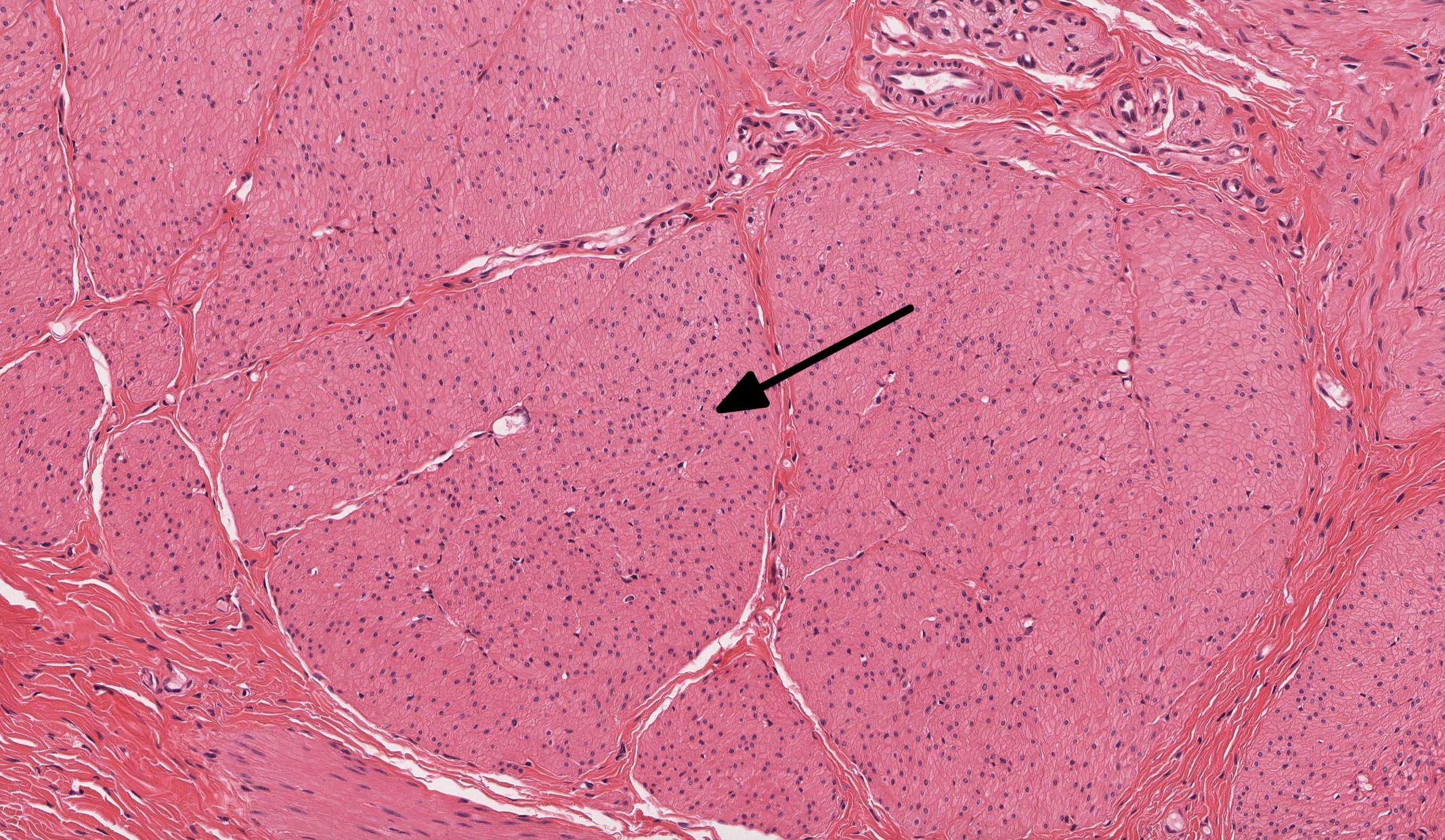
View High Mag Image
- It contracts rapidly and voluntarily.
- It interacts directly with nerves via motor end plates.
- It consists of regularly arranged sarcomeres.
- It can readily regenerate and/or proliferate via cell division.
- ALL of the above statements are true.
Answer
Correct answer 4. Can readily regenerate and/or proliferate via cell division. The tissue shown is smooth muscle; all of the other responses apply to skeletal muscle (well, choice 3 actually applies to both cardiac AND skeletal muscle).
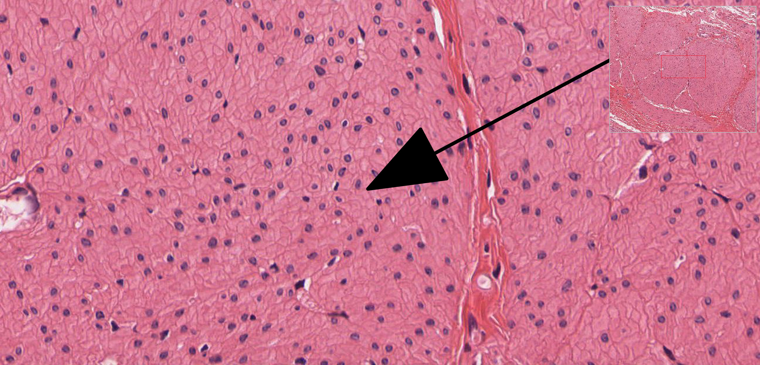
3. Identify the tissue in this micrograph.
View Image
- Skeletal muscle
- Cardiac muscle
- Smooth muscle
- Epimysium
- Dense regular connective tissue (tendon)
Answer
Correct answer 2. The tissue is cardiac muscle. Note the striations, branched fibers, intercalated discs, and centrally-located nuclei.
4. Which of the lines in this electron micrograph indicates a single sarcomere?
View Image
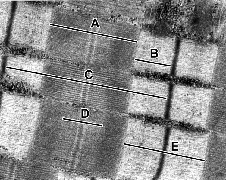
- A
- B
- C
- D
- E
Answer
Correct answer 3. (C). A sarcomere is the region spanning from one Z line to another.