1. Identify the histological components of the mammary gland, including structures associated with the nipple and the areola, the overall organization into lobes and lobules, as well as secretory alveoli (acini), lactiferous ducts/sinuses, and the intralobular and interlobular connective tissue.
2. Identify and describe the histological differences between the mammary gland in adult females prior to pregnancy (inactive), during pregnancy, and during lactation (active).
3. Know the cellular mechanisms involved in the formation and release of milk.
Slide 265 Nipple, areola H&E View Virtual Slide
The 16-20 lactiferous ducts slide 265 Nipple, areola H&E View Image, one from each lobe, open at the summit of the nipple. These ducts are lined by stratified squamous epithelium near the opening and the lumens are frequently filled with desquamated cells. Deeper in the connective tissue, the ducts acquire a stratified columnar appearance that is really a cuboidal duct cell sitting on a myoepithelial cell as in the sweat gland.
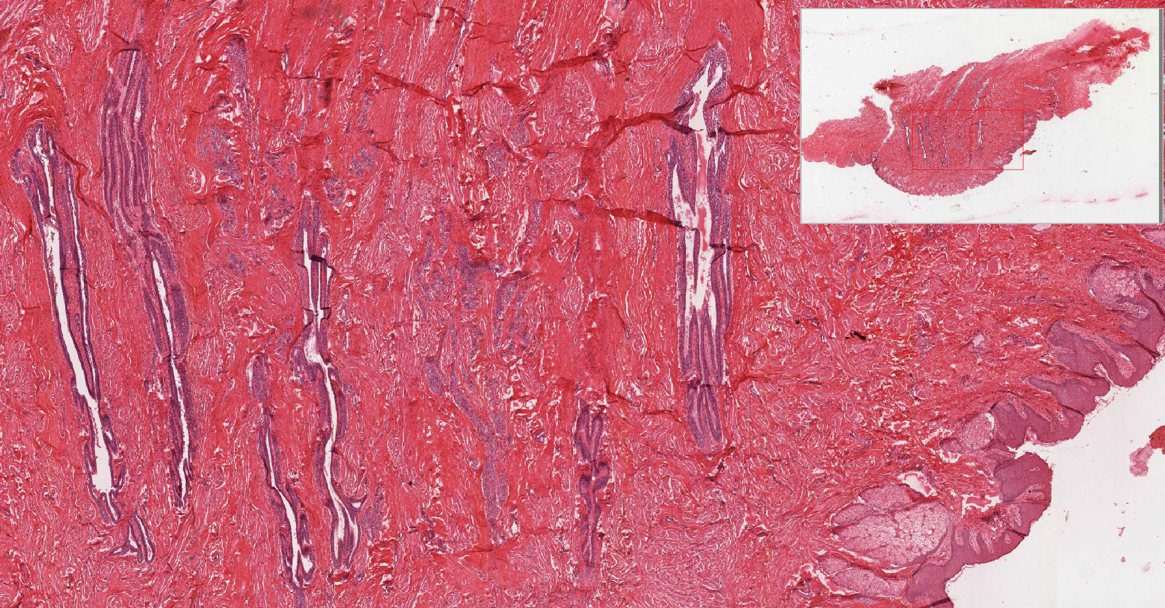
Sebaceous glands slide 265 View Image are present to a variable extent, especially in the areola. Note that the dense irregular connective tissue of the dermis is interrupted by numerous fascicles of smooth muscle slide 265 View Image that insert into the dermal connective tissue (much like arrector pili muscles). These muscle bundles are responsible for erection of the nipples. Occasional nerves are also present in the dermis.
Slide 259 Mammary gland inactive nulliparous H&E View Virtual Slide
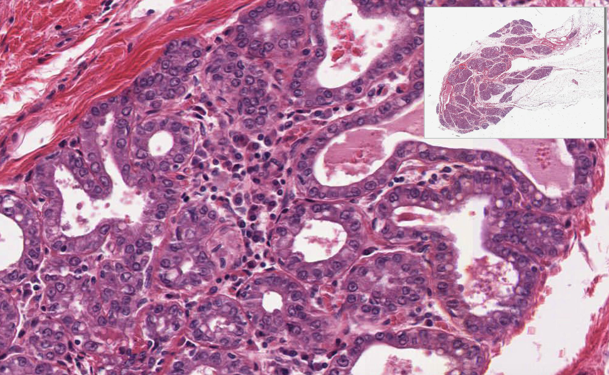
Slide 258 Mammary gland active, pregnant H&E View Virtual Slide
Slide 261 Mammary gland active lactating H&E View Virtual Slide
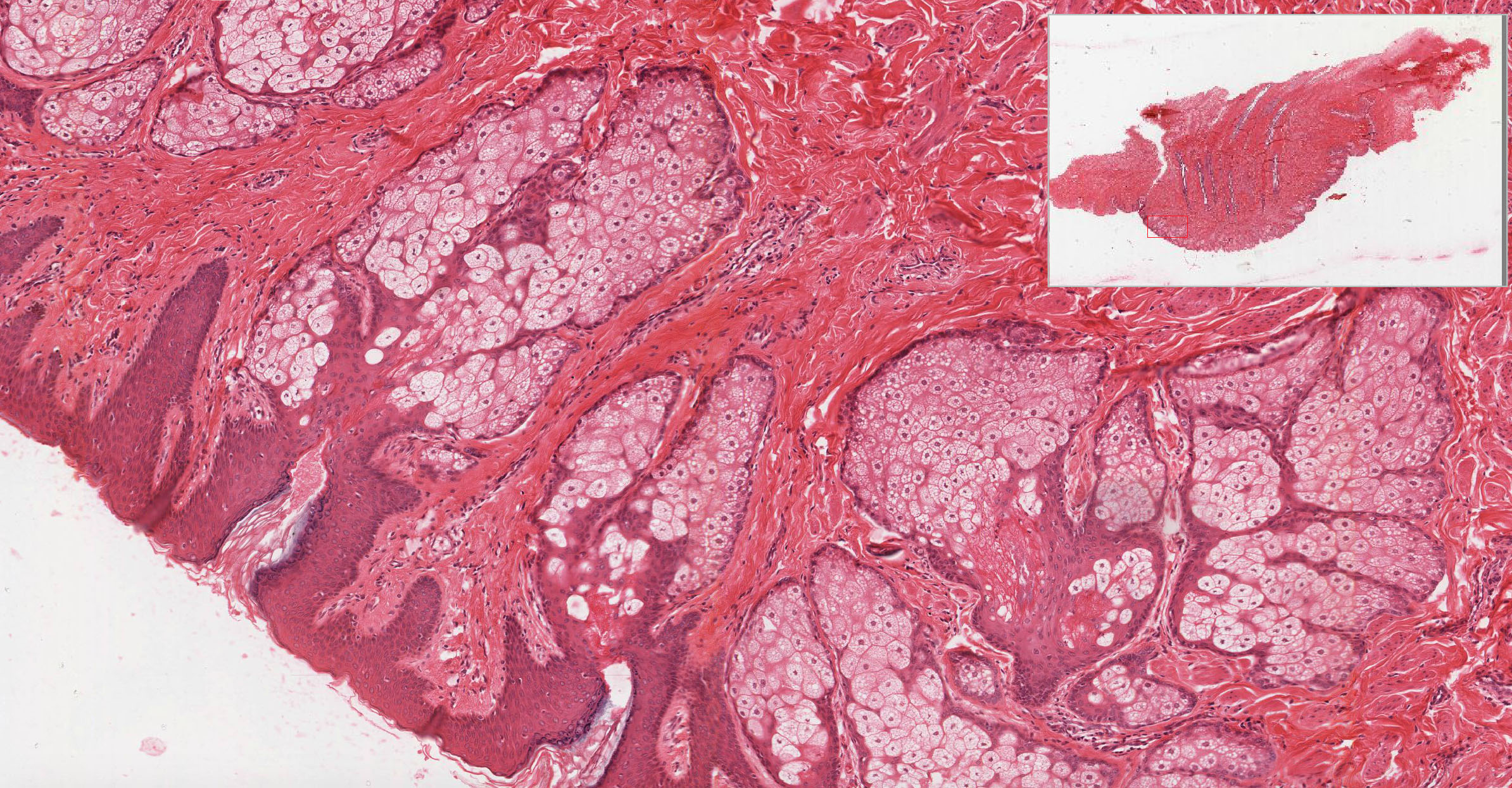
Like the other tissues in the female reproductive system, alterations in circulating hormone levels result in histologically demonstrable changes in the mammary gland. Compare the examples of an inactive and active glands, noting the differences in the amount of glandular tissues. In slide 259 (inactive gland) note the dense irrengular interlobular connective tissue found between quiescent glandular lobulesthat consist of only a few clusters of small ducts surrounded by a mass of less dense intralobular connective tissue. Many ducts appear to be composed of 2 layers of cuboidal epithelium. The inner layer are the actual ductal epithelial cells whereas the outer layer of cells is, in fact, a layer of myoepithelial cells.
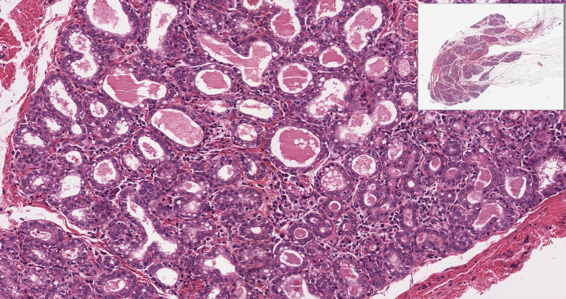
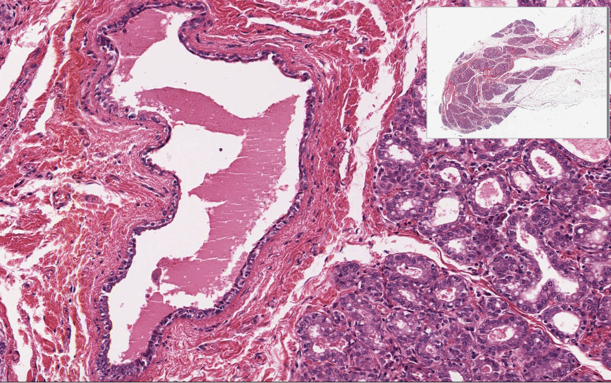
In slide 258 (active gland), you can see that the amount of the glandular tissues has increased, while that of the connective tissue has decreased. This increase involves the numbers of both the epithelial cells and myoepithelial cells. The proliferation of these cells lead to the formation of secretory alveoli. Note also the increased cellularity (especially, the plasma cells) of the intralobular connective tissue. This tissue was probably taken from an individual before the last trimester. When compared with the inactive mammary gland, you can see that the intralobular ducts have proliferated to form additional secretory regions. Both the epithelial cells and myoepithelial cells increase in number. Alveoli slide 261 View Image have formed, their epithelial cells have large, clear areas of apical cytoplasm, a region occupied by glycogen and lipid. Note the increased cellularity of the intralobular connective tissue. Note also that not all lobules within the gland have proliferated to the same degree. In many sections, portions of a large excretory, lactiferous duct slide 261 View Image are present. The epithelial lining is again two layered, the bottom layer being principally myoepithelium. Compare the morphology of the inactive and active gland. Observe the intralobular connective tissue and note the abundance of plasma cells slide 261 intralobular connective tissue and note the abundance of plasma cells View Image. These plasma cells are the source of secretory IgA.
290 Mammary gland View Virtual EM Slide
Observe the process of true apocrine secretion. Lipid granules are released covered with a small amount of mammary gland cell cytoplasm, as well as the cell membrane. On the other hand, the protein components are released by the usual mechanism of exocytosis.
Click on a question to reveal the answer.
What is the difference between intralobular and interlobular connective tissue?
Intralobular connective tissue (the connective tissue around the ducts of each lobule) is a looser, more vascular and cellular connective tissue. The cellularity comes not only from fibroblasts, but also from an abundant number of plasma cells that produce the IgA that is secreted into the milk. Interlobular (between lobules) connective tissue, on the other hand, is dense and collagenous and contains fewer cells.